Tuberc Respir Dis > Volume 74(6); 2013 > Article
Park and Yoo: The Clinical Assessment of Protease-Activated Receptor-2 Expression in Inflammatory Cells from Peripheral Blood and Bronchoalveolar Lavage Fluid in Idiopathic Pulmonary Fibrosis

Abstract

Background

Idiopathic pulmonary fibrosis (IPF) is a lethal pulmonary fibrotic disease. In general, the exaggerated activation of the coagulation cascade has been observed during initiation or maintenance of the fibrotic disease. In our recent study, immunohistochemical expression of protease-activated receptor-2 (PAR-2), which plays a key role in coagulation cascade, was observed in surgical specimen of IPF patients, and associated with poor clinical outcome. The aim of this study was to evaluate the overexpression of PAR-2 in inflammatory cells from peripheral blood and bronchoalveolar lavage fluid in IPF patients.

Methods

From May 2011 to March 2012, IPF patients and controls were enrolled in Seoul National University Hospital. Peripheral blood and bronchoalveolar lavage fluid were collected for analysis of PAR-2 expression. Flow cytometry and reverse transcription polymerase chain reaction were used for PAR-2 receptor and mRNA assessment.

Results

Twelve IPF patients and 14 controls were included in this study. Among them, flow cytometry analysis was conducted from 26 peripheral blood (patient group, 11; control group, 13) and 7 bronchoalveolar lavage fluid (patient group, 5; control group, 2). The expression of PAR-2 receptor was not different between patient and control groups (p=0.074). Among all 24 population, PAR-2 mRNA assessment was performed in 19 persons (patient group, 10; control group, 9). The mRNA expression of PAR-2 was not significant different (p=0.633).

Conclusion

In IPF patients, PAR-2 receptor and mRNA expression were not different from control group.

Introduction

Idiopathic pulmonary fibrosis (IPF) is an inflammatory fibrotic lung disease of unknown etiology. Unfortunately, IPF is a progressive and irreversible disorder. In general, median survival of IPF patients is 2.5 to 5 years with or without treatment1,2. The pathogenesis of IPF is poorly understood3. One of the major advances in the pathogenesis of IPF is the shift in current paradigms from inflammation to abnormal wound healing4. In the wound healing process, coagulation cascade is activated locally from tissue factor dependent extrinsic pathway5. Recent studies implicated protease-activated receptor-2 (PAR-2) in the fibrosis.
PAR-2 is a G-coupled 7-transmembrane receptor that is activated by tethered peptide ligand, which is exposed after enzymatic cleavage of the specific site in the extracellular N-terminal6. PAR-2 is expressed in various tissues and abundant in kidney, pancreas, and gastrointestinal tissue than in heart and lungs7. In each organ, PAR-2 expressions were mainly located in endothelial and epithelial cells8. Recent studies suggested association of PAR-2 activation with airway inflammation9-11 and pulmonary fibrosis12. In addition, up-regulation of PAR-2 was observed in lung tissue of IPF patients and possible pathway of the development of pulmonary fibrosis13. Recently we conducted PAR-2 expression of surgical specimen from IPF patients, and we found the possibility of the association between PAR-2 expression and clinical outcome of IPF14. So the aim of this study was to evaluate the overexpression of PAR-2 in inflammatory cells from peripheral blood and bronchoalveolar lavage fluid in IPF patients.

Materials and Methods

1. Study population

We included the patients with IPF in Seoul National University between May 2011 and March 2012. The diagnosis of IPF was based on the established criteria (20) and clinical diagnosis was judged according to international guidelines. Peripheral blood and bronchoalveolar lavage fluid were collected after informed consent. The protocol was approved by the Institutional Review Board of the Seoul National University Hospital. All patients gave written informed consent. The study was conducted in accordance with the Declaration of Helsinki.

2. Mononuclear cell isolation

Blood (5 mL) was drawn into heparinized tubes and mononuclear cell isolation was performed within 6 hours from acquisition of specimen. Blood was diluted phosphate buffered saline with equal volume. The mixture was layered over 5 mL Ficoll-Paque-Plus solution (density 1.077/mL; Amersham Biosciences, Uppsala, Sweden). The layers of density gradient were separated after centrifugation (2,100 rpm for 20 minutes). Mononuclear cells were harvested with micropipette.

3. Flow cytometry

Mononuclear cells (1.0×10⁶/mL) were fixed in 4% paraformaldehyde for 20 minutes at 4℃ and then incubated 30 minutes room temperature with CD3 (BD Biosciences, San Jose, CA, USA), CD14 (BD Biosciences), PAR-2 (Santa Cruz Biotechnology, Santa Cruz, CA, USA). Each isotype controls were used for calibration.

4. Reverse transcription polymerase chain reaction (RT-PCR) and PCR

The total RNA was extracted from the mononuclear cells using RNeasy Mini kit (Qiagen, Valencia, CA, USA), and PCR was followed using commercially available kit (Bioneer, Daejeon, Korea), according to manufacturere's instructions. Oligonucleotide primers (forward: 5'-GTT GAT GGC ACA TCC CAC GTC-3', reverse: 5'-GTA CAG GGC ATA GAC ATG GC-3') were designed for PAR-2. The PCR conditions were as follows; a pre-denaturating for 94℃ for 1 minute, followed by 40 cycles fo denaturation at 94℃ for 1 minute, annealing at 64℃ for 1 minute, and extension at 72℃ for 1 minute. PCR products were separated by electrophoresis through 1% agarose gel and the signal intensity was analyzed by ImageJ. Glyceraldehyde-3-phosphate dehydrogenase (GAPDH) controls were used to standardize the quantification of samples.
RT-PCR was conducted to examination the mRNA expression of PAR-2 and GAPDH.

5. Statistical analysis

All data were showed as mean±SD. Student t-test was used for comparison between two groups. SPSS version 19.0 (SPSS Inc., Chicago, IL, USA) was used for statistical analysis and p<0.05 was considered as statistically significant difference.

Results

1. Study population

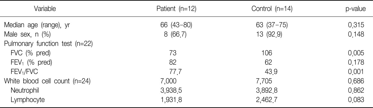
Table 1 shows the clinical characteristics of IPF patients (n=12) and controls (n=14). Median age was 66 in IPF group and 63 control group. Pulmonary function test showed statistically significant different forced vital capacity (FVC) and forced expiratory volume in one second/FVC, because chronic obstructive pulmonary disease (COPD) patients (n=8) were included in control group.

2. PAR-2 receptor expression in flow cytometry

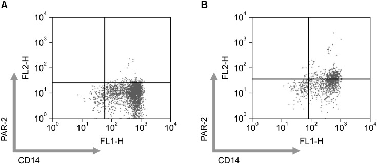
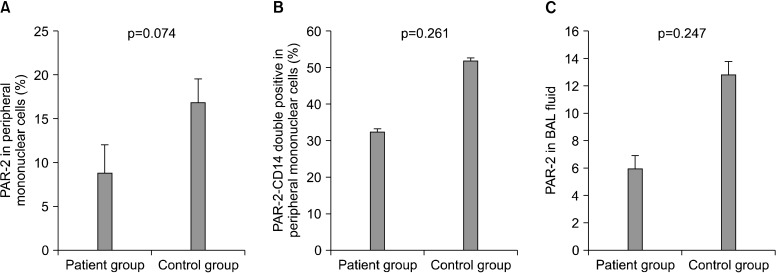
We conducted flow cytometry analysis from 26 peripheral blood (patient group, 11; control group, 13) and 7 bronchoalveolar lavage (BAL) fluid (patient group, 5; control group, 2) (Figure 1). CD3, CD14, CD20, PAR-2 expression were analyzed. PAR-2 expression was not significant different between patient and control group in peripheral blood and BAL fluid (Figure 2). The proportion of PAR-2 and CD14 double positive among CD14 single positive was no different in both groups (p=0.247).

3. PAR-2 mRNA expression

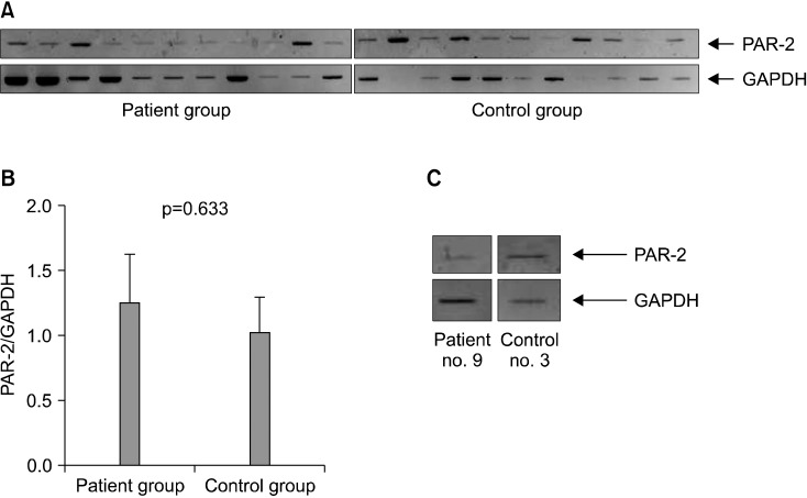
Among 26 persons, PAR-2 mRNA assessment was performed in 19 persons (patient group, 10; control group, 9). mRNA expression of PAR-2 was not different between patient and control group (p=0.633) (Figure 3). Because there was paucity of cellularity in BAL fluid, only two cases (patient, 1; control, 1) were conducted RT-PCR for mRNA expression of PAR-2.

Discussion

The understanding of IPF pathogenesis has shifted from inflammatory response to aberrant wound healing of unknown etiology4. In other words, the pulmonary fibrosis is considered as the common end pathway of aberrant wound healing. Interestingly, during the fibrotic process, coagulation cascade activation was frequently observed not only IPF but also other inflammatory conditions such as systemic sclerosis and acute respiratory distress syndrome15-17. In acute lung injury animal model, uncontrolled activation of coagulation cascade and pulmonary fibrosis was also observed, and furthermore PAR-2 activated by Factor Xa was a significant receptor for the development of pulmonary fibrosis12. These findings suggest the possible role of PAR-2 in the pathogenesis or progression of pulmonary fibrosis.
As described above, we reported the relationship between immunohistochemical expression of PAR-2 and worse clinical outcomes of IPF patients14. But this study did not show an increased PAR-2 receptor expression in BAL fluid and peripheral mononuclear cells with IPF patients. The relationship between activation of coagulation cascade and pathogenesis of IPF might be irrelevant. Moreover, warfarin did not show a beneficial effect in progressive IPF in recent study18. In addition, this study had some limitations. Most of all, study population was so small. We included only 12 IPF patients during 10 months. The second, control group was not homogeneous. Among 14 control group, 8 were COPD patients. COPD is well known systemic inflammatory disease and PAR-2 is associated with inflammation19. So it is explainable that PAR-2 receptor expression was increased in control group (Figure 3).
In conclusion, PAR-2 receptor and mRNA expression was not increased in IPF patients compared to control group.

Acknowledgements

This Study was supported by a 2011 Grant from The Korean Academy of Tuberculosis and Respiratory Diseases.

References

1. American Thoracic Society. European Respiratory Society. American Thoracic Society/European Respiratory Society International Multidisciplinary Consensus Classification of the Idiopathic Interstitial Pneumonias. This joint statement of the American Thoracic Society (ATS), and the European Respiratory Society (ERS) was adopted by the ATS board of directors, June 2001 and by the ERS Executive Committee, June 2001. Am J Respir Crit Care Med 2002;165:277-304. PMID: 11790668.
crossref pmid
2. Harari S, Caminati A. Idiopathic pulmonary fibrosis. Allergy 2005;60:421-435. PMID: 15727572.
crossref pmid
3. Caminati A, Harari S. IPF: new insight in diagnosis and prognosis. Respir Med 2010;104(Suppl 1):S2-S10. PMID: 20639137.
crossref pmid
4. Selman M, King TE, Pardo A. American Thoracic Society. European Respiratory Society. American College of Chest Physicians. Idiopathic pulmonary fibrosis: prevailing and evolving hypotheses about its pathogenesis and implications for therapy. Ann Intern Med 2001;134:136-151. PMID: 11177318.
crossref pmid
5. Chambers RC. Procoagulant signalling mechanisms in lung inflammation and fibrosis: novel opportunities for pharmacological intervention? Br J Pharmacol 2008;153(Suppl 1):S367-S378. PMID: 18223674.
crossref pmid pmc
6. Nystedt S, Emilsson K, Wahlestedt C, Sundelin J. Molecular cloning of a potential proteinase activated receptor. Proc Natl Acad Sci U S A 1994;91:9208-9212. PMID: 7937743.
crossref pmid pmc
7. Nystedt S, Emilsson K, Larsson AK, Strombeck B, Sundelin J. Molecular cloning and functional expression of the gene encoding the human proteinase-activated receptor 2. Eur J Biochem 1995;232:84-89. PMID: 7556175.
crossref pmid
8. D'Andrea MR, Derian CK, Leturcq D, Baker SM, Brunmark A, Ling P, et al. Characterization of protease-activated receptor-2 immunoreactivity in normal human tissues. J Histochem Cytochem 1998;46:157-164. PMID: 9446822.
crossref pmid
9. Miotto D, Hollenberg MD, Bunnett NW, Papi A, Braccioni F, Boschetto P, et al. Expression of protease activated receptor-2 (PAR-2) in central airways of smokers and non-smokers. Thorax 2002;57:146-151. PMID: 11828045.
crossref pmid pmc
10. Knight DA, Lim S, Scaffidi AK, Roche N, Chung KF, Stewart GA, et al. Protease-activated receptors in human airways: upregulation of PAR-2 in respiratory epithelium from patients with asthma. J Allergy Clin Immunol 2001;108:797-803. PMID: 11692107.
crossref pmid
11. Peters T, Henry PJ. Protease-activated receptors and prostaglandins in inflammatory lung disease. Br J Pharmacol 2009;158:1017-1033. PMID: 19845685.
crossref pmid pmc
12. Borensztajn K, Bresser P, van der Loos C, Bot I, van den Blink B, den Bakker MA, et al. Protease-activated receptor-2 induces myofibroblast differentiation and tissue factor up-regulation during bleomycin-induced lung injury: potential role in pulmonary fibrosis. Am J Pathol 2010;177:2753-2764. PMID: 20971733.
crossref pmid pmc
13. Wygrecka M, Kwapiszewska G, Jablonska E, von Gerlach S, Henneke I, Zakrzewicz D, et al. Role of protease-activated receptor-2 in idiopathic pulmonary fibrosis. Am J Respir Crit Care Med 2011;183:1703-1714. PMID: 21471103.
crossref pmid
14. Park YS, Park CM, Lee HJ, Goo JM, Chung DH, Lee SM, et al. Clinical implication of protease-activated receptor-2 in idiopathic pulmonary fibrosis. Respir Med 2013;107:256-262. PMID: 23131707.
crossref pmid
15. Hernandez-Rodriguez NA, Cambrey AD, Harrison NK, Chambers RC, Gray AJ, Southcott AM, et al. Role of thrombin in pulmonary fibrosis. Lancet 1995;346:1071-1073. PMID: 7564789.
crossref pmid
16. Imokawa S, Sato A, Hayakawa H, Kotani M, Urano T, Takada A. Tissue factor expression and fibrin deposition in the lungs of patients with idiopathic pulmonary fibrosis and systemic sclerosis. Am J Respir Crit Care Med 1997;156(2 Pt 1):631-636. PMID: 9279250.
crossref pmid
17. Gunther A, Mosavi P, Heinemann S, Ruppert C, Muth H, Markart P, et al. Alveolar fibrin formation caused by enhanced procoagulant and depressed fibrinolytic capacities in severe pneumonia: comparison with the acute respiratory distress syndrome. Am J Respir Crit Care Med 2000;161(2 Pt 1):454-462. PMID: 10673185.
crossref pmid
18. Noth I, Anstrom KJ, Calvert SB, de Andrade J, Flaherty KR, Glazer C, et al. A placebo-controlled randomized trial of warfarin in idiopathic pulmonary fibrosis. Am J Respir Crit Care Med 2012;186:88-95. PMID: 22561965.
crossref pmid pmc
19. Lewkowich IP, Day SB, Ledford JR, Zhou P, Dienger K, Wills-Karp M, et al. Protease-activated receptor 2 activation of myeloid dendritic cells regulates allergic airway inflammation. Respir Res 2011;12:122PMID: 21936897.
crossref pmid pmc pdf
Figure 1
Distribution of protease-activated receptor-2 (PAR-2) and CD14 double positive cells in idiopathic pulmonary fibrosis patient (A) and control (B).
trd-74-264-g001.jpg
Figure 2
Comparison of protease-activated receptor-2 (PAR-2) expression in flow cytometry between patient and control group. PAR-2 expression was not significantly different in peripheral blood (A) and bronchoalveolar lavage fluid (C). The fraction of PAR-2 and CD14 double positive population in CD14 positive population was not significant different (B).
trd-74-264-g002.jpg
Figure 3
mRNA expression in peripheral mononuclear cells (A) and bronchoalveolar lavage fluid (C). The difference of mRNA expression in peripheral mononuclear cells was not significantly different (B, p=0.633). PAR-2: protease-activated receptor-2; GAPDH: glyceraldehyde-3-phosphate dehydrogenase.
trd-74-264-g003.jpg
Table 1
Baseline characteristics of included patients with idiopathic pulmonary fibrosis and controls
trd-74-264-i001.jpg

FVC: forced vital capacity; FEV1: forced expiratory volume in one second.


ABOUT
ARTICLE & TOPICS
Article category

Browse all articles >

Topics

Browse all articles >

BROWSE ARTICLES
FOR CONTRIBUTORS
Editorial Office
101-605, 58, Banpo-daero, Seocho-gu (Seocho-dong, Seocho Art-Xi), Seoul 06652, Korea
Tel: +82-2-575-3825, +82-2-576-5347    Fax: +82-2-572-6683    E-mail: katrdsubmit@lungkorea.org                

Copyright © 2024 by The Korean Academy of Tuberculosis and Respiratory Diseases. All rights reserved.

Developed in M2PI

Close layer
prev next