Tuberc Respir Dis > Volume 87(1); 2024 > Article
Lee and Song: Diagnostic Approaches for Idiopathic Pulmonary Fibrosis

Abstract

Idiopathic pulmonary fibrosis (IPF) is a chronic, progressive, fibrosing interstitial pneumonia with a very poor prognosis. Accurate diagnosis of IPF is essential for good outcomes but remains a major medical challenge due to variability in clinical presentation and the shortcomings of existing diagnostic tests. Medical history collection is the first and most important step in the IPF diagnosis process; the clinical probability of IPF is high if the suspected patient is 60 years or older, male, and has a history of cigarette smoking. Systemic assessment for connective tissue disease is essential in the initial evaluation of patients with suspected IPF to identify potential causes of interstitial lung disease (ILD). Radiologic examination using high-resolution computed tomography plays a pivotal role in the evaluation of patients with ILD, and prone and expiratory computed tomography images can be considered. If additional tests such as surgical lung biopsy or transbronchial lung cryobiopsy are needed, transbronchial lung cryobiopsy should be considered as an alternative to surgical lung biopsy in medical centers with experience performing this procedure. Diagnosis through multidisciplinary discussion (MDD) is strongly recommended as MDD has become the cornerstone for diagnosis of IPF, and the scope of MDD has expanded to monitoring of disease progression and suggestion of appropriate treatment options.

Key Figure

Introduction

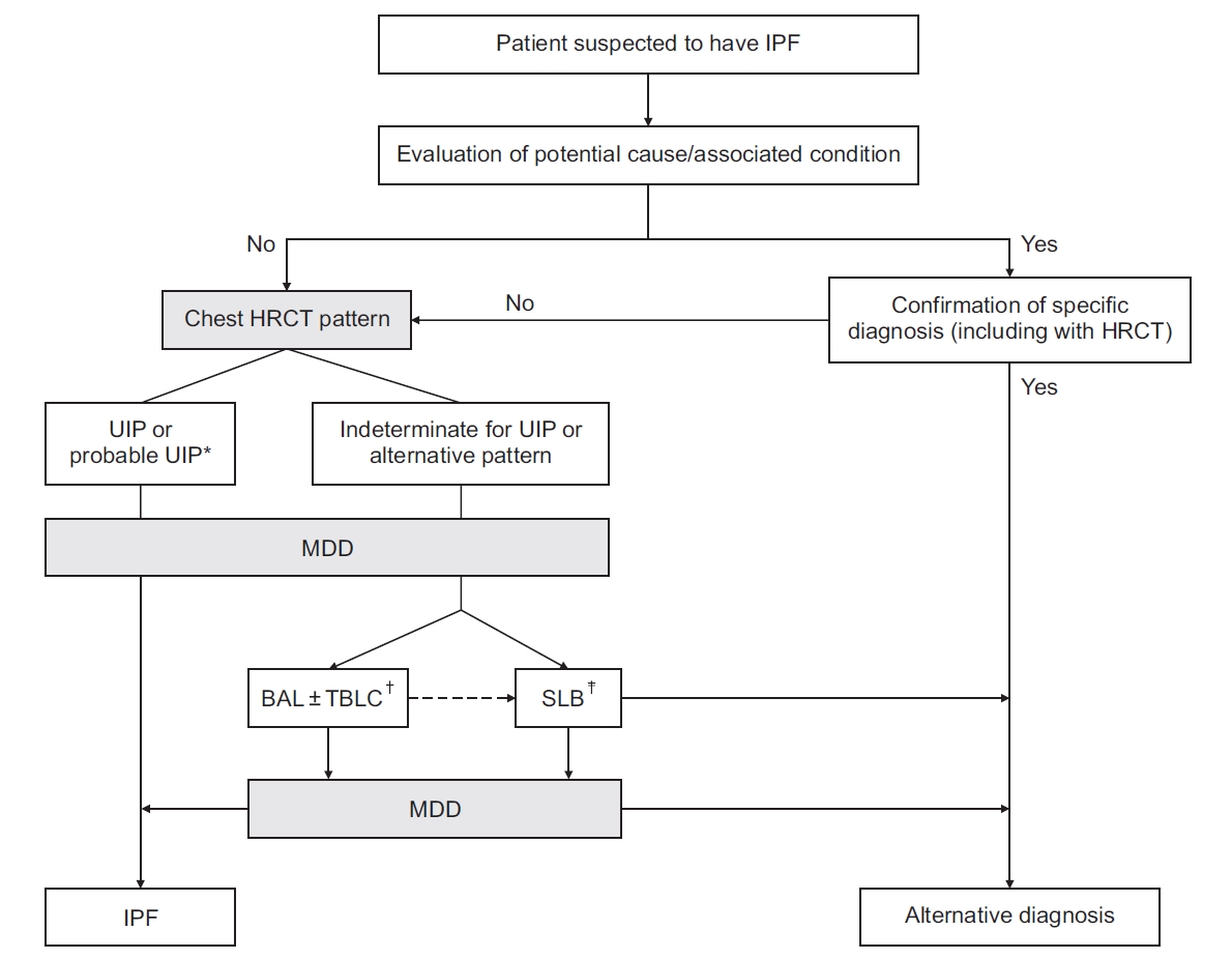
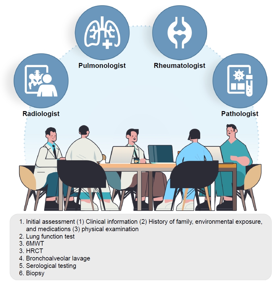
Idiopathic pulmonary fibrosis (IPF) is a chronic, progressive, fibrosing interstitial pneumonia characterized by progression and poor prognosis, with a median survival of 3 to 5 years [1-3]. Therefore, accurate diagnosis is essential to optimize early treatment for better outcomes in IPF [4,5]. However, accurate diagnosis of IPF remains challenging due to variability in the interpretation of radiopathologic findings and the shortcomings of existing diagnostic tests 6 . Since the first IPF international guidelines were published in 2001, diagnosis of IPF has evolved through a better understanding of the disease mechanism and advances in diagnostic methods including high-resolution computed tomography (HRCT), biopsy techniques, and multidisciplinary discussion (MDD) [7]. Currently, diagnosis of IPF is based on the dynamic integration of clinical, radiologic, and histopathologic information through MDD (Figure 1). The aim of this review is to address the current diagnostic process for IPF based on emerging evidence and the most recent international guidelines.

Initial Assessment

Collection of medical history is the first and most important step in the IPF diagnosis process. If interstitial lung disease (ILD) is clinically suspected and identified on HRCT, focused history collection about medication use and environmental exposure at home, work, and other frequently visited places in addition to physical examination should be performed to exclude potential causes of ILD [8]. The clinical probability of IPF is high if the patient is 60 years or older, male, and has a history of cigarette smoking 9 . Common clinical features of patients with IPF are shortness of breath, dry cough, and fatigue. However, these clinical symptoms are nonspecific and are often attributed to other conditions such as airway disease, cancer, and heart disease.
In terms of physical examination, bibasilar inspiratory crackles are commonly detected on chest auscultation, and finger clubbing is present in approximately 50% of patients with IPF [10,11]. Lung function parameters including forced vital capacity, total lung capacity (TLC), and diffusion capacity for carbon monoxide are usually reduced but may be normal in early or subclinical IPF stages. The 6-minute walk test (6MWT) is a reliable tool for practical assessment of functional exercise capacity to determine overall cardiopulmonary reserve [12]. Assessment of total distance and arterial oxygen saturation at the beginning and end of the 6MWT should be measured and used as predictors of mortality [12-15].

Radiologic Assessment

Radiologic examination using HRCT plays a pivotal role in the evaluation of patients with ILD and is routinely performed to diagnose IPF. The Fleischner Society and American Thoracic Society (ATS)/ European Respiratory Society (ERS)/ Japanese Respiratory Society (JRS)/ Latin American Thoracic Association (ALAT) guidelines outline the technical requirements for HRCT to obtain optimal-quality computed tomography (CT) images [16,17]. Thin sections (<2 mm), short rotation times, and high spatial resolution reconstruction are needed. Images should be obtained at full inspiration to TLC [18,19]. Volumetric CT acquisition is preferred over non-contiguous imaging to improve the characterization of patch disease and to delineate the extent of disease. Prone CT images may be considered due to the resolution of atelectasis in the prone position, when there is minimal abnormal opacification of dependent opacification on supine CT images [20]. Prone CT images can enhance the detection of honeycombing and reduce observer variation in IPF diagnosis [21]. Expiratory images can be considered to identify air trapping, which would suggest an alternative diagnosis such as fibrotic hypersensitivity pneumonitis [22].

HRCT Patterns

The ATS/ERS/JRS/ALAT guidelines specify four diagnostic HRCT patterns: usual interstitial pneumonia (UIP), probable UIP, indeterminate UIP, and alternative diagnosis (Table 1) [17].
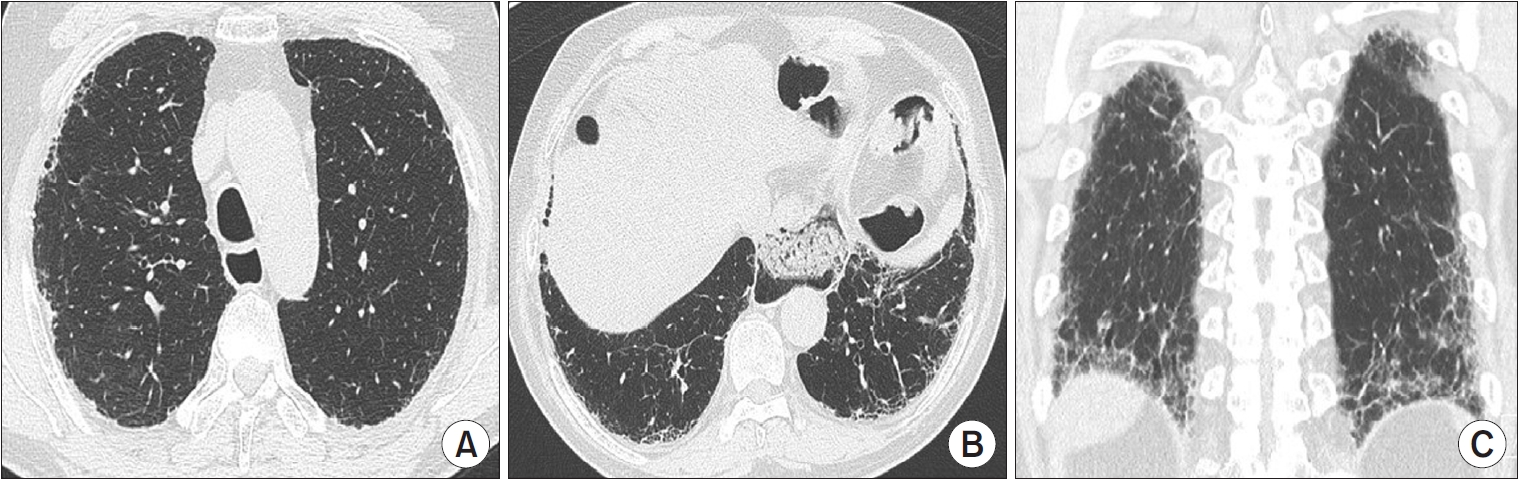
A UIP pattern is the characteristic radiological pattern of IPF. Honeycombing is a necessary and distinguishing feature of a UIP pattern on HRCT (Figure 2). It may be present with or without peripheral traction bronchiectasis or bronchiolectasis. The typical distribution of UIP is subpleural with basal predominance. In a minority of cases, the cranio-caudal distribution of UIP may be relatively uniform, and asymmetric disease may occur in up to 25% of cases [23,24]. In a previous study that investigated the likelihood of histologic confirmation of UIP in patients with a UIP pattern on HRCT, the confidence level of a radiologic UIP pattern for UIP histology was between 90% and 100% [25,26]. Patients with a UIP pattern on HRCT can be diagnosed with IPF without the need for a surgical lung biopsy (SLB) after exclusion of other causes.
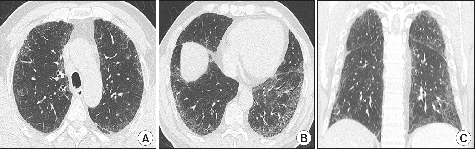
The “possible UIP” category in the 2011 guidelines was updated to “probable UIP” in the 2018 international guidelines because studies showed that selected patients with a “possible UIP” pattern on HRCT are highly likely to have UIP histology, even without evidence of honeycombing on HRCT [27]. Reticular abnormalities with traction bronchiectasis or bronchiolectasis with a subpleural and basal predominant distribution are defined as “probable UIP” (Figure 3). Level of confidence for UIP histology is between 70% and 89% [27-29]. Among patients with a probable UIP pattern on HRCT, those with clinical risk factors (age 60 years or older, male, cigarette smokers) can be diagnosed with IPF after MDD without confirmation by lung biopsy.
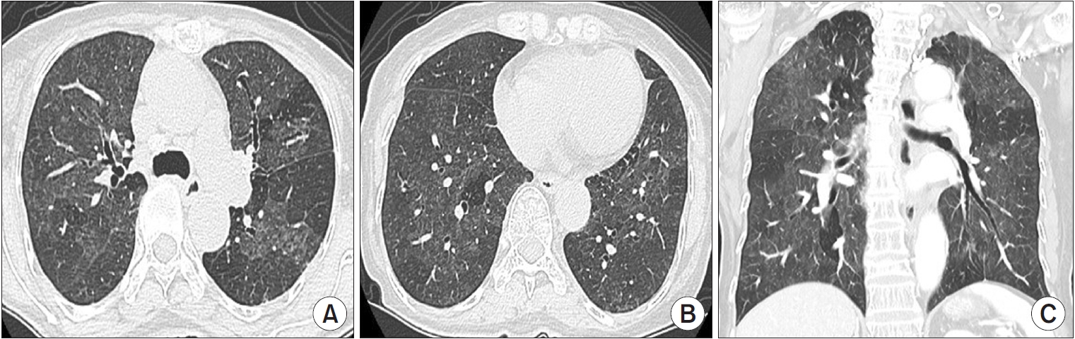
“Indeterminate for UIP pattern” is assigned when HRCT demonstrates features of lung fibrosis that do not suggest any specific diagnosis (Figure 4). In the revised 2022 ATS/ERS/JRS/ALAT guidelines, this category includes CT features of lung fibrosis with a diffuse distribution and without subpleural predominance. The level of confidence for UIP histology is between 51% and 69% [30-32].
Alternative diagnosis pattern on HRCT suggests a diagnosis other than IPF (Figure 5). This may include bronchocentric fibrosis in the upper lobes, profuse mosaic attenuation suggesting hypersensitivity pneumonitis, or extensive ground-glass opacification with subpleural sparing in nonspecific interstitial pneumonia (NSIP). The level of confidence for UIP histology is less than 50% [30,31].

Rheumatologic Assessment

Systemic assessment for connective tissue disease (CTD) is essential in the initial evaluation of patients with suspected IPF to identify potential causes of ILD. Although there is no clear consensus, autoantibody serologic panels, C-reactive protein, erythrocyte sedimentation rate, antinuclear antibodies, rheumatoid factor, anti-cyclic citrullinated peptide, myositis panel, and muscle enzymes should be evaluated. Current international guidelines recommend that patients be referred for rheumatologic evaluation if they have extrapulmonary disease manifestations suggesting CTD and serologic abnormalities or other characteristics not consistent with IPF (middle-aged, females, non-smokers, or family history of ILD) [17]. In some patients, lung manifestations are the first or only dominant feature of CTD. Some patients also might not fulfil the standard rheumatologic diagnostic criteria at the time of ILD diagnosis, but some of them may be newly diagnosed with CTD during follow-up [33,34]. Therefore, follow-up by a rheumatologist needs to be considered in these patients [34-36].

Bronchoalveolar Lavage

Bronchoalveolar lavage (BAL) is a procedure performed through flexible bronchoscopy to obtain a sample of alveolar cells. BAL findings are usually considered nonspecific for ILD. The percentage of neutrophils in IPF is higher (range, 5.9% to 22.08%) than that in healthy individuals (≤3%) or patients with other ILDs [17,37]. Patients with IPF have a higher proportion of macrophages than those with other ILDs, but the macrophage proportion is lower in individuals with ILDs than healthy individuals (>85%). In the 2011 guidelines, cellular analysis of BAL fluid was not recommended for diagnostic evaluation of IPF due to the additional risk and cost [38]. However, in the 2018 guidelines, BAL fluid cellular analysis is recommended for patients clinically suspected of IPF with an HRCT pattern of probable UIP, indeterminate for UIP, or an alternative diagnosis (conditional recommendation, very low quality of evidence); cellular analysis is not recommended for individuals with a UIP pattern [17,30]. Cellular analysis of BAL fluid may be useful in distinguishing IPF from some alternative ILDs such as eosinophilic pneumonia (eosinophil percentage ≥25%), hypersensitivity pneumonitis (lymphocyte percentage ≥30%), and sarcoidosis (lymphocyte percentage ≥15% and CD4 to CD8 ratio >4:1) [30,39,40].

Histopathologic Examination

UIP is a histopathologic term introduced in 1969 as part of the initial classification of interstitial pneumonia. Histopathologic hallmarks of UIP are the microscopic features of patch dense fibrosis associated with remodeling of lung architecture resulting in honeycomb changes alternating with areas of less affected parenchyma with a mainly subpleural and paraseptal distribution. IPF diagnosis has been based on the histopathologic pattern of UIP; however, there are concerns regarding the risks of SLB and the high frequency of interobserver disagreement in histologic analysis [41]. All patients with suspected IPF do not need surgical biopsy, and the final decision regarding whether to perform this surgery must consider the risks and benefits for individual patients.

Histopathologic Patterns

There are four diagnostic categories of IPF based on HRCT pattern: UIP, probable UIP, indeterminate UIP, and alternative diagnosis (Table 2) [17]. A UIP pattern is defined as patchy, dense fibrosis with the following features: (1) remodeling of lung architecture; (2) honeycomb changes; and (3) distribution that alternates with area of less affected parenchyma, typically in subpleural and paraseptal areas. Probable UIP has some but not all histopathologic features of UIP; features to suggest an alternative diagnosis are absent or only honeycombing may be present. Indeterminate for UIP is defined as fibrosis with or without architectural distortion, favoring either a pattern other than UIP or UIP secondary to another cause. Alternative diagnosis has histologic patterns of idiopathic interstitial pneumonia without fibroblastic foci or loose fibrosis in all biopsies in addition to histologic features indicative of other diseases.

Surgical Lung Biopsy

SLB is the gold standard method for histopathological diagnosis of patients with ILD and is indicated when a non-invasive diagnosis is not possible. About one-third of IPF patients in previous studies and clinical trials were diagnosed using SLB [3-5,42]. This method yields adequate specimens for histopathologic diagnosis with a median diagnostic yield around 95% [43]. However, SLB is associated with procedure-related mortality (1.7% and 16% for elective and non-elective SLB), respiratory infections, and delayed wound healing [41]. Definite diagnosis can be obtained in about 90% of patients, but there is a high frequency of interobserver disagreement regarding histopathologic diagnosis [44,45]. Use of SLB to diagnose IPF has decreased over time [46]. However, SLB remains an important diagnostic test to determine adequate treatment through accurate diagnosis in certain patients with ILD.

Transbronchial Lung Cryobiopsy

SLB, including open lung biopsy or video-assisted thoracoscopic lung biopsy, increases patient burden, but transbronchial lung biopsy may not be optimal for diagnosis of IPF because of a small sample size or imaging artifacts [47]. Transbronchial lung cryobiopsy (TBLC) is a new diagnostic approach for diffuse ILDs that is increasingly being used to diagnose IPF. Accumulation of experience over decades and technological advancements have improved diagnostic yield and safety profiles. Recently, the diagnostic yield was reported to be 79%, which is higher than that reported in a previous meta-analysis (72.9%), and can be as high as 85% when three or more samples are collected [48-52]. In terms of complications, reported rates of pneumothorax and pneumothorax requiring chest tube drainage are 9% and 5.6%, respectively, and other severe complications including severe bleeding (1.6%), procedural mortality (0.6%), and exacerbations (1.4%) are uncommon [53-56]. The improved safety profiles compared to previous studies (pneumothorax incidence, 12%; 95% CI, 3% to 21% and moderate/severe bleeding incidence, 39%; 95% CI, 3% to 76%) may be due to accumulation of experience and preventive interventions including endobronchial balloon blockers and fluoroscopic guidance [57]. In the 2020 guidelines of the American College of Chest Physicians and the 2022 ATS/ERS guidelines, TBLC is recommended as an acceptable alternative approach to SLB for histopathologic diagnosis in patients with probable UIP, indeterminate for UIP, or alternative diagnosis on HRCT. Procedural risks, patient preference for TBLC versus SLB, and the experience of the medical center with performing and interpreting TBLC should be considered when deciding to perform TBLC [30,58].

Multidisciplinary Discussion

Given the broad differential diagnosis for IPF, accurate diagnosis is difficult but crucial because of the prognostic and therapeutic implications. Since the joint statement on classification of idiopathic interstitial pneumonia of ATS/ERS in 2002, MDD involving a pulmonologist, a radiologist, and a histopathologist is recommended to diagnose ILD [59]. Evidence in favor of a multidisciplinary approach integrating clinical, radiologic, and histopathologic information has been increasing [60,61]. Walsh et al. [62] investigated 70 patients with ILD and reported that the inter-MDD agreement on diagnosis of IPF (weight-kappa coefficient [kw]=0.71 [interquartile range (IQR), 0.64 to 0.77]) was better than that for idiopathic non-NSIP (kw=0.42 [IQR, 0.37 to 0.49]) and hypersensitivity pneumonitis (kw=0.29 [IQR, 0.24 to 0.40]), and MDD resulted in a diagnosis of IPF with high confidence (77%). Based on these findings, the authors suggested that MDD could also improve interobserver agreement between MDDs for diagnosis of IPF [62]. Therefore, recent guidelines emphasize the importance of dynamic assessment by MDD, and MDD has become the cornerstone for diagnosis and management of ILD [30,36,38]. Despite MDD, however, some patients are not diagnosed and classified with a specific ILD. Therefore, efforts are being made to present confidence levels as a provisional diagnosis in ILD classification during MDD [63]. Originally, MDD was developed and implemented to improve the diagnostic accuracy of ILD, but the scope of MDD has expanded to monitoring of disease progression and determining optimal treatments to achieve better outcomes [64]. Through monitoring of disease progression, MDD can propose changes in treatment including participation in clinical trials, timely implementation of non-pharmacological therapies (supplemental oxygen or pulmonary rehabilitation), and preparation for lung transplantation, resulting in a better prognosis.

Conclusion

Accurate diagnosis of IPF remains a major medical challenge due to variability in clinical presentation and limitations of current diagnostic tests. For better outcomes, a detailed medical history including systemic assessment for connective tissue disease is the first and most important step. Radiologic examination using HRCT plays a pivotal role in the evaluation of patients with ILD. If histopathologic samples are needed, SLB or TBLC may be considered. MDD has become the cornerstone of diagnosis of IPF, and the scope of MDD now includes monitoring of disease progression and suggestion of appropriate treatment options.

Notes

Authors’ Contributions

Conceptualization: Song JW. Methodology: Song JW. Formal analysis: all authors. Data curation: Lee JH. Software: all authors. Validation: Song JW. Writing - original draft preparation: Lee JH. Writing - review and editing: all authors. Approval of final manuscript: all authors.

Conflicts of Interest

Jin Woo Song is an editorial board member of the journal, but he was not involved in the peer reviewer selection, evaluation, or decision process of this article. No other potential conflicts of interest.

Funding

This study was supported by grants from the Basic Science Research Program (NRF-2022R1A2B5B0200 1602) and the Bio & Medical Technology Development Program (NRF-2022M3A9E4082647) of the National Research Foundation of Korea (NRF) funded by the Ministry of Science & ICT, Republic of Korea, as well as by grants from the National Institute of Health research project (2021ER120701) and the Korea Environment Industry & Technology Institute through the Core Technology Development Project for Environmental Diseases Prevention and Management Program funded by the Korea Ministry of the Environment (ARQ202201450001), Republic of Korea.

Figure 1.
Diagnostic algorithm for idiopathic pulmonary fibrosis (IPF). Adopted from Raghu et al. [17] *Patients with a radiological pattern of probable usual interstitial pneumonia can be diagnosed with IPF after multidisciplinary discussion without confirmation by lung biopsy in the appropriate clinical setting (i.e., 60 years or older, male, smoker). Transbronchial lung cryobiopsy may be preferred to surgical lung biopsy in centers with appropriate expertise and/ or in some patient populations. Surgical lung biopsy may be justified in some patients with nondiagnostic finding on transbronchial lung cryobiopsy. HRCT: high-resolution computed tomography; UIP: usual interstitial pneumonia; MDD: multidisciplinary discussion; BAL: bronchoalveolar lavage; TBLC: transbronchial lung cryobiopsy; SLB: surgical lung biopsy.
trd-2023-0087f1.jpg
Figure 2.
Usual interstitial pneumonia pattern on high-resolution computed tomography (HRCT) images. Axial HRCT scans at the upper (A) and the lower (B) lung zones show subpleural reticulation and honeycombing mainly in the lower lobes. Coronal reformation image (C) demonstrates the peripheral distribution of fibrosis and the basal predominance of honeycombing with traction bronchiectasis.
trd-2023-0087f2.jpg
Figure 3.
Probable usual interstitial pneumonia pattern on high-resolution computed tomography (HRCT) images. Axial HRCT scans at the upper lobes (A) and the lung bases (B) show a bilateral subpleural reticular pattern, which is more severe in the basal lungs. Note the large esophageal hiatal hernia. Coronal reformation image (C) shows subpleural and basal predominant intralobular lines and irregular septal thickening resulting in a reticular pattern, which is associated with traction bronchiolectasis and also mild traction bronchiectasis.
trd-2023-0087f3.jpg
Figure 4.
Indeterminate for usual interstitial pneumonia (UIP) pattern on high-resolution computed tomography (HRCT) images. Axial (A, B) and coronal (C) reformation HRCT scans at the upper (A) and lower (B) lung zones show diffuse mild to moderate ground-glass opacities and fine reticulation with no subpleural or craniocaudal predominance. These computed tomography features of lung fibrosis are not suggestive of any specific diagnosis.
trd-2023-0087f4.jpg
Figure 5.
Alternative diagnosis on high-resolution computed tomography (HRCT) images. Axial (A, B) and coronal (C) reformation HRCT scans at the upper (A) and lower (B) lung zones show diffuse ground-glass opacity with poorly defined centrilobular micronodules interposed with areas of normal lung and lobular areas of decreased attenuation. The combination of ground-glass opacity, normal lung, and areas of decreased attenuation gives the lung an appearance suggestive of hypersensitivity pneumonitis.
trd-2023-0087f5.jpg
trd-2023-0087f6.jpg
Table 1.
High-resolution computed tomography patterns
HRCT pattern
UIP pattern Probable UIP pattern Indeterminate UIP Alternative diagnosis
Level of confidence for UIP histology Confident (>90%) Provisional high confidence (70%-89%) Provisional low confidence (51%-69%) Low to very low confidence (≤50%)
Distribution Subpleural and basal predominant Subpleural and basal predominant Diffuse distribution without subpleural predominance Peribronchovascular predominant with subpleural sparing (consider NSIP)
Often heterogeneous (areas of normal lung interspersed with fibrosis) Often heterogeneous (areas of normal lung interspersed with reticulation and traction bronchiectasis/ bronchiolectasis) Perilymphatic distribution (consider sarcoidosis)
Occasionally diffuse Upper or mid lung (consider fibrotic HP, CTDILD, and sarcoidosis)
May be asymmetric Subpleural sparing (consider NSIP or smoking-related IP)
CT features Honeycombing with or without traction bronchiectasis/bronchiolectasis Reticular pattern with traction bronchiectasis/bronchiolectasis CT features of lung fibrosis that do not suggest any specific etiology Lung findings
Presence of irregular thickening of interlobular septa  Cysts (consider LAM, PLCH, LIP, and DIP)
Usually superimposed with a reticular pattern and mild GGO May have mild GGO  Mosaic attenuation or three-density sign (consider HP)
May have pulmonary ossification Absence of subpleural sparing  Predominant GGO (consider HP, smokingrelated disease, drug toxicity, and acute exacerbation of fibrosis)
 Profuse centrilobular micronodules (consider HP or smoking-related disease)
 Nodules (consider sarcoidosis)
 Consolidation (consider organizing pneumonia, etc.)
Mediastinal findings
 Pleural plaques (consider asbestosis)
 Dilated esophagus (consider CTD)

Adopted from Raghu et al. [17]

HRCT: high-resolution computed tomography; UIP: usual interstitial pneumonia; NSIP: nonspecific interstitial pneumonia; HP: hypersensitivity pneumonitis; CTD-ILD: connective tissue disease-associated interstitial lung disease; IP: interstitial pneumonia; CT: computed tomography; GGO: ground-glass opacity; LAM: lymphangioleiomyomatosis; PLCH: pulmonary Langerhans cell histiocytosis; LIP: lymphoid interstitial pneumonia; DIP: desquamative interstitial pneumonia; CTD: connective tissue disease.

Table 2.
Histopathology patterns and features
UIP Probable UIP Indeterminate for UIP Alternative diagnosis
Dense fibrosis with architectural distortion (i.e., destructive scarring and/or honeycombing) Some histologic features from column 1 are present but to an extent that precludes a definite diagnosis of UIP/IPF Fibrosis with or without architectural distortion, with features favoring either a pattern other than UIP or UIP secondary to another cause* Features of other histologic patterns of IIPs (e.g., absence of fibroblast foci or predominant subpleural and/or loose fibrosis) in all biopsies
Predominant subpleural and/or paraseptal distribution of fibrosis And
Patchy involvement of lung parenchyma by fibrosis Absence of features to suggest an alternative diagnosis Some histologic features from column 1 but with other features suggesting an alternative diagnosis Histologic findings indicative of other diseases (e.g., hypersensitivity pneumonitis, Langerhans cell histiocytosis, sarcoidosis, LAM)
Fibroblast foci Or
Absence of features to suggest an alternate diagnosis Honeycombing only

Adopted from Raghu et al. [17]

* Granulomas, hyaline membranes (other than when associated with acute exacerbation of IPF, which may be the presenting manifestation in some patients), prominent airway-centered changes, areas of interstitial inflammation lacking associated fibrosis, marked chronic fibrous pleuritis, organizing pneumonia.

Features that should raise concerns about the likelihood of an alternative diagnosis include a cellular inflammatory infiltrate away from areas of honeycombing, prominent lymphoid hyperplasia including secondary germinal centers, and a distinctly bronchiolocentric distribution that could include extensive peribronchiolar metaplasia.

UIP: usual interstitial pneumonia; IPF: idiopathic pulmonary fibrosis; IIP: idiopathic interstitial pneumonia; LAM: lymphangioleiomyomatosis.

REFERENCES

1. Ley B, Collard HR, King TE Jr. Clinical course and prediction of survival in idiopathic pulmonary fibrosis. Am J Respir Crit Care Med 2011;183:431-40.
crossref pmid
2. du Bois RM, Weycker D, Albera C, Bradford WZ, Costabel U, Kartashov A, et al. Ascertainment of individual risk of mortality for patients with idiopathic pulmonary fibrosis. Am J Respir Crit Care Med 2011;184:459-66.
crossref pmid
3. Fernandez Perez ER, Daniels CE, Schroeder DR, St Sauver J, Hartman TE, Bartholmai BJ, et al. Incidence, prevalence, and clinical course of idiopathic pulmonary fibrosis: a population-based study. Chest 2010;137:129-37.
crossref pmid pmc
4. Richeldi L, du Bois RM, Raghu G, Azuma A, Brown KK, Costabel U, et al. Efficacy and safety of nintedanib in idiopathic pulmonary fibrosis. N Engl J Med 2014;370:2071-82.
pmid
5. King TE Jr, Bradford WZ, Castro-Bernardini S, Fagan EA, Glaspole I, Glassberg MK, et al. A phase 3 trial of pirfenidone in patients with idiopathic pulmonary fibrosis. N Engl J Med 2014;370:2083-92.
crossref pmid
6. Wells AU, Costabel U, Poletti V, Crestani B, Egan J, Margaritopoulos G, et al. Challenges in IPF diagnosis, current management and future perspectives. Sarcoidosis Vasc Diffuse Lung Dis 2015;32 Suppl 1:28-35.
pmid
7. American Thoracic Society. Idiopathic pulmonary fibrosis: diagnosis and treatment. International consensus statement. American Thoracic Society (ATS), and the European Respiratory Society (ERS). Am J Respir Crit Care Med 2000;161(2 Pt 1):646-64.
crossref
8. Singh S, Collins BF, Sharma BB, Joshi JM, Talwar D, Katiyar S, et al. Interstitial lung disease in India: results of a prospective registry. Am J Respir Crit Care Med 2017;195:801-13.
pmid
9. Brownell R, Moua T, Henry TS, Elicker BM, White D, Vittinghoff E, et al. The use of pretest probability increases the value of high-resolution CT in diagnosing usual interstitial pneumonia. Thorax 2017;72:424-9.
crossref pmid pmc
10. Wuyts WA, Dahlqvist C, Slabbynck H, Schlesser M, Gusbin N, Compere C, et al. Baseline clinical characteristics, comorbidities and prescribed medication in a real-world population of patients with idiopathic pulmonary fibrosis: the PROOF registry. BMJ Open Respir Res 2018;5:e000331.
crossref pmid pmc
11. Behr J, Kreuter M, Hoeper MM, Wirtz H, Klotsche J, Koschel D, et al. Management of patients with idiopathic pulmonary fibrosis in clinical practice: the INSIGHTS-IPF registry. Eur Respir J 2015;46:186-96.
crossref pmid pmc
12. ATS Committee on Proficiency Standards for Clinical Pulmonary Function Laboratories. ATS statement: guidelines for the six-minute walk test. Am J Respir Crit Care Med 2002;166:111-7.
crossref pmid
13. Gupta R, Ruppel GL, Espiritu JR. Exercise-induced oxygen desaturation during the 6-minute walk test. Med Sci (Basel) 2020;8:8.
crossref pmid pmc
14. Jenkins S, Cecins N. Six-minute walk test: observed adverse events and oxygen desaturation in a large cohort of patients with chronic lung disease. Intern Med J 2011;41:416-22.
crossref pmid
15. Lama VN, Flaherty KR, Toews GB, Colby TV, Travis WD, Long Q, et al. Prognostic value of desaturation during a 6-minute walk test in idiopathic interstitial pneumonia. Am J Respir Crit Care Med 2003;168:1084-90.
crossref pmid
16. Lynch DA, Sverzellati N, Travis WD, Brown KK, Colby TV, Galvin JR, et al. Diagnostic criteria for idiopathic pulmonary fibrosis: a Fleischner Society White Paper. Lancet Respir Med 2018;6:138-53.
crossref pmid
17. Raghu G, Remy-Jardin M, Myers JL, Richeldi L, Ryerson CJ, Lederer DJ, et al. Diagnosis of idiopathic pulmonary fibrosis: an official ATS/ERS/JRS/ALAT clinical practice guideline. Am J Respir Crit Care Med 2018;198:e44-68.
pmid
18. Mayo JR. CT evaluation of diffuse infiltrative lung disease: dose considerations and optimal technique. J Thorac Imaging 2009;24:252-9.
pmid
19. Bankier AA, O’Donnell CR, Boiselle PM. Quality initiatives: respiratory instructions for CT examinations of the lungs: a hands-on guide. Radiographics 2008;28:919-31.
crossref pmid
20. Hansell DM. Thin-section CT of the lungs: the Hinterland of normal. Radiology 2010;256:695-711.
crossref pmid
21. Kim M, Lee SM, Song JW, Do KH, Lee HJ, Lim S, et al. Added value of prone CT in the assessment of honeycombing and classification of usual interstitial pneumonia pattern. Eur J Radiol 2017;91:66-70.
crossref pmid
22. Tokura S, Okuma T, Akira M, Arai T, Inoue Y, Kitaichi M. Utility of expiratory thin-section CT for fibrotic interstitial pneumonia. Acta Radiol 2014;55:1050-5.
crossref pmid pdf
23. Gruden JF, Panse PM, Leslie KO, Tazelaar HD, Colby TV. UIP diagnosed at surgical lung biopsy, 2000-2009: HRCT patterns and proposed classification system. AJR Am J Roentgenol 2013;200:W458-67.
crossref pmid
24. Tcherakian C, Cottin V, Brillet PY, Freynet O, Naggara N, Carton Z, et al. Progression of idiopathic pulmonary fibrosis: lessons from asymmetrical disease. Thorax 2011;66:226-31.
crossref pmid
25. Hunninghake GW, Zimmerman MB, Schwartz DA, King TE Jr, Lynch J, Hegele R, et al. Utility of a lung biopsy for the diagnosis of idiopathic pulmonary fibrosis. Am J Respir Crit Care Med 2001;164:193-6.
crossref pmid
26. Nishimura K, Izumi T, Kitaichi M, Nagai S, Itoh H. The diagnostic accuracy of high-resolution computed tomography in diffuse infiltrative lung diseases. Chest 1993;104:1149-55.
crossref pmid
27. Chung JH, Chawla A, Peljto AL, Cool CD, Groshong SD, Talbert JL, et al. CT scan findings of probable usual interstitial pneumonitis have a high predictive value for histologic usual interstitial pneumonitis. Chest 2015;147:450-9.
crossref pmid pmc
28. Fukihara J, Kondoh Y, Brown KK, Kimura T, Kataoka K, Matsuda T, et al. Probable usual interstitial pneumonia pattern on chest CT: is it sufficient for a diagnosis of idiopathic pulmonary fibrosis? Eur Respir J 2020;55:1802465.
crossref pmid
29. Chung JH, Oldham JM, Montner SM, Vij R, Adegunsoye A, Husain AN, et al. CT-pathologic correlation of major types of pulmonary fibrosis: insights for revisions to current guidelines. AJR Am J Roentgenol 2018;210:1034-41.
crossref pmid
30. Raghu G, Remy-Jardin M, Richeldi L, Thomson CC, Inoue Y, Johkoh T, et al. Idiopathic pulmonary fibrosis (an update) and progressive pulmonary fibrosis in adults: an official ATS/ERS/JRS/ALAT clinical practice guideline. Am J Respir Crit Care Med 2022;205:e18-47.
pmid pmc
31. Sumikawa H, Johkoh T, Colby TV, Ichikado K, Suga M, Taniguchi H, et al. Computed tomography findings in pathological usual interstitial pneumonia: relationship to survival. Am J Respir Crit Care Med 2008;177:433-9.
crossref pmid
32. Flaherty KR, Thwaite EL, Kazerooni EA, Gross BH, Toews GB, Colby TV, et al. Radiological versus histological diagnosis in UIP and NSIP: survival implications. Thorax 2003;58:143-8.
crossref pmid pmc
33. Fernandes L, Nasser M, Ahmad K, Cottin V. Interstitial pneumonia with autoimmune features (IPAF). Front Med (Lausanne) 2019;6:209.
crossref pmid pmc
34. Fischer A, Antoniou KM, Brown KK, Cadranel J, Corte TJ, du Bois RM, et al. An official European Respiratory Society/American Thoracic Society research statement: interstitial pneumonia with autoimmune features. Eur Respir J 2015;46:976-87.
crossref pmid
35. Levi Y, Israeli-Shani L, Kuchuk M, Epstein Shochet G, Koslow M, Shitrit D. Rheumatological assessment is important for interstitial lung disease diagnosis. J Rheumatol 2018;45:1509-14.
crossref pmid
36. Lee SH, Yeo Y, Kim TH, Lee HL, Lee JH, Park YB, et al. Korean guidelines for diagnosis and management of interstitial lung diseases: part 2. Idiopathic pulmonary fibrosis. Tuberc Respir Dis (Seoul) 2019;82:102-17.
crossref pmid pmc pdf
37. Meyer KC, Raghu G, Baughman RP, Brown KK, Costabel U, du Bois RM, et al. An official American Thoracic Society clinical practice guideline: the clinical utility of bronchoalveolar lavage cellular analysis in interstitial lung disease. Am J Respir Crit Care Med 2012;185:1004-14.
crossref pmid
38. Raghu G, Collard HR, Egan JJ, Martinez FJ, Behr J, Brown KK, et al. An official ATS/ERS/JRS/ALAT statement: idiopathic pulmonary fibrosis: evidence-based guidelines for diagnosis and management. Am J Respir Crit Care Med 2011;183:788-824.
pmid pmc
39. Raghu G, Remy-Jardin M, Ryerson CJ, Myers JL, Kreuter M, Vasakova M, et al. Diagnosis of hypersensitivity pneumonitis in adults: an official ATS/JRS/ALAT clinical practice guideline. Am J Respir Crit Care Med 2020;202:e36-69.
pmid pmc
40. Thillai M, Atkins CP, Crawshaw A, Hart SP, Ho LP, Kouranos V, et al. BTS clinical statement on pulmonary sarcoidosis. Thorax 2021;76:4-20.
crossref pmid
41. Hutchinson JP, Fogarty AW, McKeever TM, Hubbard RB. In-hospital mortality after surgical lung biopsy for interstitial lung disease in the United States: 2000 to 2011. Am J Respir Crit Care Med 2016;193:1161-7.
crossref pmid
42. Ferrara G, Carlson L, Palm A, Einarsson J, Olivesten C, Skold M. Idiopathic pulmonary fibrosis in Sweden: report from the first year of activity of the Swedish IPF-Registry. Eur Clin Respir J 2016;3:31090.
crossref pmid pmc
43. Nguyen W, Meyer KC. Surgical lung biopsy for the diagnosis of interstitial lung disease: a review of the literature and recommendations for optimizing safety and efficacy. Sarcoidosis Vasc Diffuse Lung Dis 2013;30:3-16.
pmid
44. Nicholson AG, Fulford LG, Colby TV, du Bois RM, Hansell DM, Wells AU. The relationship between individual histologic features and disease progression in idiopathic pulmonary fibrosis. Am J Respir Crit Care Med 2002;166:173-7.
crossref pmid
45. Hutchinson J, Hubbard R, Raghu G. Surgical lung biopsy for interstitial lung disease: when considered necessary, should these be done in larger and experienced centres only? Eur Respir J 2019;53:1900023.
crossref pmid
46. Eldersveld JM, Yi ES, Kunze KL, Smith ML, Tazelaar HD, Larsen BT. Usual interstitial pneumonia in contemporary surgical pathology practice: impact of International Consensus Guidelines for Idiopathic Pulmonary Fibrosis on Pathologists. Arch Pathol Lab Med 2021;145:717-27.
crossref pmid pdf
47. Kebbe J, Abdo T. Interstitial lung disease: the diagnostic role of bronchoscopy. J Thorac Dis 2017;9(Suppl 10):S996-1010.
crossref pmid pmc
48. Troy LK, Grainge C, Corte TJ, Williamson JP, Vallely MP, Cooper WA, et al. Diagnostic accuracy of transbronchial lung cryobiopsy for interstitial lung disease diagnosis (COLDICE): a prospective, comparative study. Lancet Respir Med 2020;8:171-81.
pmid
49. Unterman A, Wand O, Fridel L, Edelstein E, Pertzov B, Kramer MR. High diagnostic accuracy of transbronchial cryobiopsy in fibrotic interstitial lung diseases compared to final explant diagnosis. Respiration 2019;98:421-7.
crossref pmid pdf
50. Ravaglia C, Wells AU, Tomassetti S, Gurioli C, Gurioli C, Dubini A, et al. Diagnostic yield and risk/benefit analysis of trans-bronchial lung cryobiopsy in diffuse parenchymal lung diseases: a large cohort of 699 patients. BMC Pulm Med 2019;19:16.
crossref pmid pmc pdf
51. Ravaglia C, Wells AU, Tomassetti S, Dubini A, Cavazza A, Piciucchi S, et al. Transbronchial lung cryobiopsy in diffuse parenchymal lung disease: comparison between biopsy from 1 segment and biopsy from 2 segments: diagnostic yield and complications. Respiration 2017;93:285-92.
crossref pmid pdf
52. Sethi J, Ali MS, Mohananey D, Nanchal R, Maldonado F, Musani A. Are transbronchial cryobiopsies ready for prime time?: a systematic review and meta-analysis. J Bronchology Interv Pulmonol 2019;26:22-32.
pmid
53. Jacob M, Bastos HN, Mota PC, Melo N, Cunha R, Pereira JM, et al. Diagnostic yield and safety of transbronchial cryobiopsy in sarcoidosis. ERJ Open Res 2019;5:00203-2019.
crossref pmid pmc
54. Ussavarungsi K, Kern RM, Roden AC, Ryu JH, Edell ES. Transbronchial cryobiopsy in diffuse parenchymal lung disease: retrospective analysis of 74 cases. Chest 2017;151:400-8.
crossref pmid
55. Pajares V, Puzo C, Castillo D, Lerma E, Montero MA, Ramos-Barbon D, et al. Diagnostic yield of transbronchial cryobiopsy in interstitial lung disease: a randomized trial. Respirology 2014;19:900-6.
crossref pmid
56. Rodrigues I, Estevao Gomes R, Coutinho LM, Rego MT, Machado F, Morais A, et al. Diagnostic yield and safety of transbronchial lung cryobiopsy and surgical lung biopsy in interstitial lung diseases: a systematic review and meta-analysis. Eur Respir Rev 2022;31:210280.
crossref pmid pmc
57. Johannson KA, Marcoux VS, Ronksley PE, Ryerson CJ. Diagnostic yield and complications of transbronchial lung cryobiopsy for interstitial lung disease: a systematic review and metaanalysis. Ann Am Thorac Soc 2016;13:1828-38.
crossref pmid
58. Maldonado F, Danoff SK, Wells AU, Colby TV, Ryu JH, Liberman M, et al. Transbronchial cryobiopsy for the diagnosis of interstitial lung diseases: CHEST Guideline and Expert Panel Report. Chest 2020;157:1030-42.
crossref pmid
59. American Thoracic Society; European Respiratory Society. American Thoracic Society/European Respiratory Society international multidisciplinary consensus classification of the idiopathic interstitial pneumonias. This joint statement of the American Thoracic Society (ATS), and the European Respiratory Society (ERS) was adopted by the ATS board of directors, June 2001 and by the ERS Executive Committee, June 2001. Am J Respir Crit Care Med 2002;165:277-304.
crossref pmid
60. Wang W, Xu J, Liu C, Feng R, Zhao J, Gao N, et al. The significance of multidisciplinary classifications based on transbronchial pathology in possible idiopathic interstitial pneumonias. Medicine (Baltimore) 2020;99:e20930.
crossref pmid pmc
61. De Sadeleer LJ, Meert C, Yserbyt J, Slabbynck H, Verschakelen JA, Verbeken EK, et al. Diagnostic ability of a dynamic multidisciplinary discussion in interstitial lung diseases: a retrospective observational study of 938 cases. Chest 2018;153:1416-23.
crossref pmid
62. Walsh SLF, Wells AU, Desai SR, Poletti V, Piciucchi S, Dubini A, et al. Multicentre evaluation of multidisciplinary team meeting agreement on diagnosis in diffuse parenchymal lung disease: a case-cohort study. Lancet Respir Med 2016;4:557-65.
pmid
63. Ryerson CJ, Corte TJ, Lee JS, Richeldi L, Walsh SL, Myers JL, et al. A standardized diagnostic ontology for fibrotic interstitial lung disease: an International Working Group Perspective. Am J Respir Crit Care Med 2017;196:1249-54.
crossref pmid pmc
64. Cottin V, Martinez FJ, Smith V, Walsh SL. Multidisciplinary teams in the clinical care of fibrotic interstitial lung disease: current perspectives. Eur Respir Rev 2022;31:220003.
crossref pmc


ABOUT
ARTICLE & TOPICS
Article category

Browse all articles >

Topics

Browse all articles >

BROWSE ARTICLES
FOR CONTRIBUTORS
Editorial Office
101-605, 58, Banpo-daero, Seocho-gu (Seocho-dong, Seocho Art-Xi), Seoul 06652, Korea
Tel: +82-2-575-3825, +82-2-576-5347    Fax: +82-2-572-6683    E-mail: katrdsubmit@lungkorea.org                

Copyright © 2024 by The Korean Academy of Tuberculosis and Respiratory Diseases. All rights reserved.

Developed in M2PI

Close layer
prev next