


Introduction
Chronic obstructive pulmonary disease (COPD) is characterized by irreversible airflow obstruction and persistent airway inflammation caused by the inhalation of noxious particles, commonly from cigarette smoking [1]. COPD is a heterogeneous condition, as the pathophysiological and clinical manifestations vary considerably between individuals [2]. For example, the presence and severity of emphysema shows marked variability between individuals. This heterogeneity causes different responses to pharmacological interventions.
Inhaled corticosteroids (ICS) are anti-inflammatory drugs that are commonly used to treat COPD patients [3]. Randomized controlled trials (RCTs) have shown that ICS combined with a long-acting β2-agonist (LABA) reduce exacerbation rates and improve both lung function and quality of life compared to LABA monotherapy [4]. Furthermore, triple therapy consisting of ICS plus LABA plus long-acting muscarinic antagonist (LAMA) in a single inhaler also demonstrates these clinical benefits compared to LAMA/LABA combination treatment or LAMA monotherapy [5-8]. These RCTs were conducted in patients with a history of exacerbations; these individuals have an increased future risk of exacerbations, as the past exacerbation history is the best predictor of future risk [9].
It is well recognized in clinical practice that the beneficial effects of ICS vary between patients [3]. Furthermore, long term ICS treatment has the potential for adverse effects, including osteoporosis, pneumonia, and cataracts [2]. While RCTs show an overall benefit for ICS on a population basis (in patients with a history of exacerbations) [4], clinical practice requires an individualized approach to the use of these drugs, in order to identify individuals more likely to gain benefit while also limiting the potential for harm [2,3].
Precision medicine combines individual clinical and biological information to enable a more personalized approach to pharmacological treatment, with the aim of identifying patients most likely to benefit while also minimizing the risk of causing harm [2]. Blood eosinophil counts have emerged as a COPD biomarker that can be combined with clinical information to enable a precision medicine approach to the use of ICS in clinical practice. This review will focus on the evidence supporting blood eosinophils as a biomarker to guide ICS use in COPD patients, and discuss practical issues regarding implementation in clinical practice.
Eosinophilic Inflammation in COPD
A number of cytokines and chemokines, including interleukin 5 (IL-5), control the maturation, trafficking, and activity of eosinophils [10]. Eosinophils secrete various proteins that promote inflammation and tissue remodeling. Studies have reported that COPD patients have increased numbers of eosinophils in sputum samples, broncho-alveolar lavage, and bronchial biopsies compared to healthy controls [11,12]; close inspection of the data shows that a subset of COPD patients have increased eosinophil numbers, while the remainder have levels similar to controls. Blood eosinophil numbers in COPD patients are also higher than age-matched healthy controls, even when patients with a history of asthma or atopy are excluded [13].
Kolsum et al. [14] reported that COPD patients with higher blood and lung eosinophil counts showed numerous other pathological differences, including increased levels of biomarkers of type 2 (T2) inflammation and greater reticular basement membrane thickening. These features are also seen in patients with asthma [15,16], but Kolsum et al. [14] carefully excluded individuals with a history of asthma or atopy, so it would be incorrect to apply an asthma label to these COPD patients with eosinophilic inflammation. Furthermore, a study comparing COPD patients with a confirmed childhood history of asthma versus COPD patients with increased eosinophils and no history of asthma showed that the former group had more evidence of allergy and more exacerbations while displaying less eosinophilic inflammation (data shown in Table 1) [17]. These data highlight that the terms âasthmaâ and âeosinophilicâ should not be used interchangeably in COPD patients.
Relationship between Blood and Lung Eosinophil Numbers
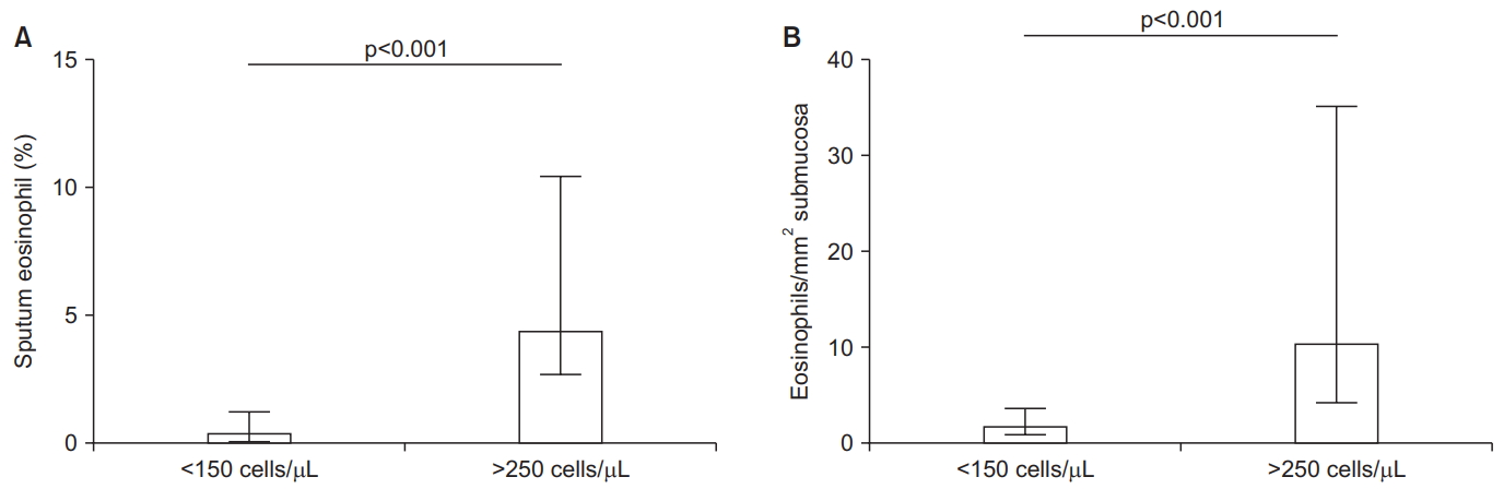
Many studies have reported statistically significant associations between blood and sputum eosinophil counts, with weak to moderate correlation coefficients (0.17-0.54) [18-22]. Factors that may negatively impact this relationship are poor quality sputum slides, probably more common in multicentre studies, and using only one significant figure for blood eosinophil counts. Studies of blood and lung eosinophil counts have shown diverse results, with both positive associations and no relationship [14,23,24]. Again, technical factors affecting lung eosinophil measurements may reduce the ability to observe an association. Nevertheless, Kolsum et al. [14] showed clearly that COPD patients with blood eosinophil counts <150 cells/ÂľL had lower bronchial mucosa, broncho-alveolar lavage, and sputum eosinophil numbers compared to COPD patients with blood eosinophil counts >250 cells/ÂľL (Figure 1). Overall, most of these studies have shown associations between blood and pulmonary eosinophil counts, indicating that blood eosinophils are a biomarker that reflects the degree of eosinophilic lung inflammation.
Modeling the Relationship between Blood Eosinophils and ICS Effects
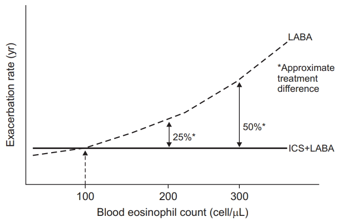
RCTs using induced sputum eosinophil counts demonstrated that COPD patients with more eosinophils had a greater forced expiratory volume in 1 second (FEV1) improvement after corticosteroid treatment [25,26]. Sputum sampling for eosinophil counts is not widely available or practical in clinical practice. Post-hoc analyses of RCTs including only COPD patients with a history of exacerbations were performed to evaluate the ability of blood eosinophil counts to predict ICS effects [27-29]. The effect of ICS/LABA treatment compared to LABA monotherapy on exacerbation prevention was greater in patients with higher blood eosinophil counts at the start of the study [27-29]. Data modeling (the INCONTROL study; n=4,528) showed that the effect of ICS treatment was observed at above approximately 100 eosinophils/ÂľL, with increasingly larger benefits at higher eosinophil counts [27]; this continuous relationship is described by Figure 2.
An important consideration for biomarkers in clinical practice is whether the results split the population in a binomial manner; this is the case for diagnostic biomarkers where a binomial categorization of âdiseaseâ or âno diseaseâ is required. In contrast, pharmacological treatment responses form a continuous spectrum (i.e., ranging from no response to small response to large response). A biomarker for predicting drug responses should therefore predict different magnitudes of response. Using blood eosinophil counts to predict ICS ârespondersâ and ânon-respondersâ is a simplistic approach that does not mirror the range of clinical responses observed. Instead, the INCONTROL data modeling results show that blood eosinophil counts can be used to predict different magnitudes of response, reflecting the population distribution of drug responses [27].
Pre-specified analysis of triple therapy studies conducted in COPD patients with a history of exacerbations have also reported greater ICS effects on exacerbation prevention in patients with higher blood eosinophil counts. In the IMPACT study (n=10,333), data modeling showed that the benefit of triple therapy compared to LABA/LAMA on exacerbation prevent was observed at above approximately 100 eosinophils/ÂľL [30]. Again, the magnitude of benefit increased at higher blood eosinophil counts, with approximately 50% exacerbation rate reduction observed at >300 eosinophils/ÂľL, as shown in Figure 2. Interestingly, ICS benefits were lower in current smokers, with ICS benefits in this subgroup observed at a higher threshold, approximately >200 eosinophils/ÂľL. A similar negative influence of current smoking on ICS effects was reported in the INCONTROL data modeling comparing ICS/LABA versus LABA [27]. The reduced effects of ICS in current smokers has not been consistently reported in COPD clinical trials and may be related to insufficient statistical power in previous subgroup analysis.
The TRIBUTE study compared triple therapy versus LABA/LAMA [7], while the TRINITY study compared triple therapy versus LAMA [6]. These studies were conducted in patients with a history of exacerbations, and in both studies it was demonstrated that a single eosinophil threshold distinguished between patients with higher and lower ICS responses e.g., in TRINITY, eosinophils âĽ2% or âĽ200 cells/ÂľL split the population into groups with approximately >30% and â¤10% exacerbation rate reductions above and below these thresholds respectively. As already discussed, these single thresholds are not the optimum way to analyze the data. The KRONOS study evaluated triple therapy in a COPD population that included patients with and without a history of exacerbations [31]. Data modeling again showed the âcontinuousâ relationship between blood eosinophil counts and ICS response, with no benefit observed at lower eosinophil counts and increasingly larger benefits at higher eosinophil counts.
Blood Eosinophils, Exacerbation History, and ICS Response
Two large studies have compared ICS/LABA versus LABA/LAMA in patients with a history of exacerbations, with different outcomes on the prevention of moderate to severe exacerbations in the overall population; in the IMPACT study, ICS/LABA had a greater effect than LABA/LAMA (10% mean difference) [8], while the FLAME study reported that LAMA/LABA had a greater effect than ICS/LABA (17% mean difference) [32]. IMPACT data modeling showed a greater effect of ICS/LABA (compared to LAMA/LABA) at higher blood eosinophil counts [30], while in the FLAME study the treatments appeared similar at higher blood eosinophil counts [33]. These studies enrolled populations with different levels of exacerbation risk; IMPACT included more patients with âĽ2 moderate exacerbations or âĽ1 severe exacerbation (hospitalization) in the previous year. This key difference appeared to increase the ICS effect (in patients with higher exacerbation risk) in IMPACT [34]. The predictive ability of blood eosinophils when comparing double combination inhalers therefore changes according to the exacerbation risk i.e., at higher eosinophil counts, any benefit of ICS/LABA over LAMA/LABA is more likely to be observed in patients at higher exacerbation risk. Other differences in the study designs of IMPACT and FLAME have been discussed and debated, such as differences in the run-in periods; previous treatment was continued during the run-in in IMPACT, while LAMA monotherapy was used in FLAME [3,34]. This led to ICS withdrawal in some patients at randomization in IMPACT but before run-in for all patients in FLAME. Regardless, the results of IMPACT and FLAME indicate that exacerbation risk and blood eosinophil counts interact to determine ICS response.
ICS withdrawal studies have demonstrated that exacerbations rates are greater in patients with higher blood eosinophil counts [35-37]. Furthermore, higher eosinophil counts plus a history of âĽ2 exacerbations appears to identify individuals who are at the greatest risk of exacerbation after ICS withdrawal [37]. The concept of ICS withdrawal in clinical practice has gained popularity in recent years, due to concerns about ICS side effects and a confidence that many patients can be treated successfully with LABA/LAMA combination inhalers without the need for additional ICS treatment. However, these ICS withdrawal studies indicate that this strategy has increased risk in patients with higher blood eosinophil counts (>300 cells/ÂľL) [3]. Furthermore, these ICS withdrawal studies were performed mainly in patients with 0 or 1 exacerbation in the previous year [35,38], but in the subgroup with âĽ2 exacerbations there appeared to be the greatest risk [37]. Again, these data highlight that blood eosinophil counts and exacerbation risk both influence ICS response.
Real word data analysis, using UK primary care information, reported that the effectiveness of triple therapy compared to LABA/LAMA was greater in patients with more exacerbations or higher blood eosinophils [39]. These real world data compliment the RCT data already reviewed.
Blood Eosinophil Count Stability
The intra-class correlation coefficient (ICC) for repeated blood eosinophil counts in COPD patients has been reported to be 0.64-0.89 in studies with follow up ranging from 3 months to 5 years [40-44]. ICC values >0.75 are interpreted as showing excellent correlation [41]. On one hand, it has been noted that these ICC values are similar to other biomarkers used in clinical practice such as cholesterol or glycated hemoglobin [3], supporting the case for using blood eosinophil counts in clinical practice. On the other hand, concerns have been expressed that blood eosinophil counts may show excessive variability, limiting their clinical usefulness [45].
It is worth dissecting published data in detail in order to understand eosinophil stability properly. A recent publication investigating stability (n=225) using the categories <100, 100 to <300 and âĽ300 eosinophils/ÂľL showed that 69.3% of COPD patients remained in the same category after 1 year [41]. Importantly, movement from one category to an adjacent category was more likely in patients with eosinophil measurements close to the threshold value, suggesting that movement between categories was more related to natural measurement variation rather than altered disease pathophysiology. GOLD 2020 recommends the use of <100 cells/ÎźL and >300 cells/ÎźL thresholds, but cautions that these are âestimates, rather than precise cut-off valuesâ for predicting ICS effects [46]. Previous criticisms of blood eosinophil count stability have focused on the re-categorization of patients due to movement across thresholds [45], but a more practical approach (as advocated by GOLD) is needed when the numerical changes are small; for example, moving from just below to just above the 300 eosinophils/ÂľL threshold does not change the clinical interpretation (namely, that there is increased likelihood of ICS benefit).
This one year stability study [41] and a different study with follow up from 2-5 years (n=59) [40] both showed that the repeatability coefficients were lower (i.e., less numerical variation) in patients with lower blood eosinophil counts. Small changes in these patients may cause movement across a threshold value (e.g., 100 eosinophils/ÂľL). However, as already discussed, it is important to understand that the clinical prediction of ICS response is unlikely to be altered by a small numerical change. Individuals with higher blood eosinophil counts (>300 cells/ÎźL) have more numerical variation, but again this may not result in any change in clinical interpretation, e.g., a change from 500 to 250 cells/ÎźL still suggests that the patient is more likely to derive benefit from ICS treatment.
Studies of blood eosinophil stability have used various thresholds, with some studies using percentage eosinophil counts rather than absolute numbers [21]. Percentage counts are clearly influenced by the presence of other immune cells, and the field has now moved towards using absolute numbers to more accurately define the degree of eosinophilic inflammation.
Blood Eosinophils and Clinical Outcomes
Numerous cohort studies have investigated associations between blood eosinophil counts and clinical features or outcomes [18,21,45,47-49]. In particular, there has been interest in whether blood eosinophil counts are associated with either exacerbation rates or mortality. The results of these studies have been inconsistent, and it is reasonable to conclude that blood eosinophil counts should not be routinely used in clinical practice as a prognostic biomarker for events such as exacerbations and mortality.
The RCTs already reviewed that were conducted in COPD patients with a history of exacerbations showed an association between higher blood eosinophil counts and increased exacerbation rates in the treatment arms without ICS (Figure 2) [27-30]. These data suggest that blood eosinophil are a prognostic biomarker in patients with both (1) a history of exacerbations and (2) receiving no ICS treatment. These RCTs showed no relationship between blood eosinophil counts and exacerbation rates in patients treated with ICS, as these drugs modify exacerbation risk in an eosinophil dependent manner. These observations provide the explanation for the lack of association between blood eosinophil counts and exacerbations in observational cohort studies, which include many patients with (1) no prior exacerbation history and/or (2) taking ICS treatment. Despite these limiting factors, some large cohort studies have still found that patients with a history of âĽ2 exacerbations plus higher blood eosinophil counts have more exacerbations at during follow-up [47].
Blood Eosinophils, Type 2 Inflammation, and Microbiome
The mechanistic explanation for the association between higher blood eosinophil counts and increased ICS response in COPD patients has not been definitively elucidated. Nevertheless, there are pieces of evidence that provide insights. The bronchoscopy study by Kolsum et al. [14] showed that higher blood eosinophil counts are associated with increased eosinophilic airway inflammation, greater reticular basement membrane thickening, and increased levels of the T2 cytokines IL-5 and eotaxin-2; these pathophysiological features are also found in patients with asthma [50,51]. However, this study excluded individuals with a history of asthma or atopy. It has also been shown that T2 gene expression in COPD bronchial biopsies is associated with lung and blood eosinophil counts [52]. Overall, eosinophilic airway inflammation seems to be associated with a wider profile of T2 inflammation; T2 inflammation is known to be corticosteroid sensitive in asthma [53], and probably the same situation exists in COPD patients.
The asthma-COPD overlap (ACO) describes a group of individuals with clinical features found in both conditions [54,55]. While âeosinophilic COPDâ shares some pathological features with asthma, there is no added value in labeling these patients as ACO, as ACO is a broad label encompassing different clinical phenotypes. Referring to these patients as âeosinophilic COPDâ or âCOPD with higher blood eosinophil countsâ is a more precise description of this COPD subset than using ACO which also includes (multiple) other subtypes.
It has been observed that higher sputum eosinophil counts are associated with lower levels of colonizing bacteria in the airways of COPD patients [56,57]. Furthermore, a longitudinal observational study reported that blood eosinophil counts <100 cells/ÂľL were associated with increased probability of chronic bacterial airway infection and pneumonia [58]. These findings suggest that susceptibility to bacterial airway infection is increased in COPD patients with lower eosinophil counts, but the mechanisms to explain these findings have not been defined [59]. Eosinophils have no direct anti-bacterial activity against common pathogens that infect COPD patients [60]. Therefore, it is likely that there are other differences in antimicrobial host defence associated with eosinophil numbers. Interestingly, low sputum eosinophil counts in COPD patients have been associated with reduced bacterial diversity and increased Proteobacteria including haemophilus [61,62]. Overall, these studies show both increased bacterial load and altered microbiome profiles in patients with lower eosinophil counts.
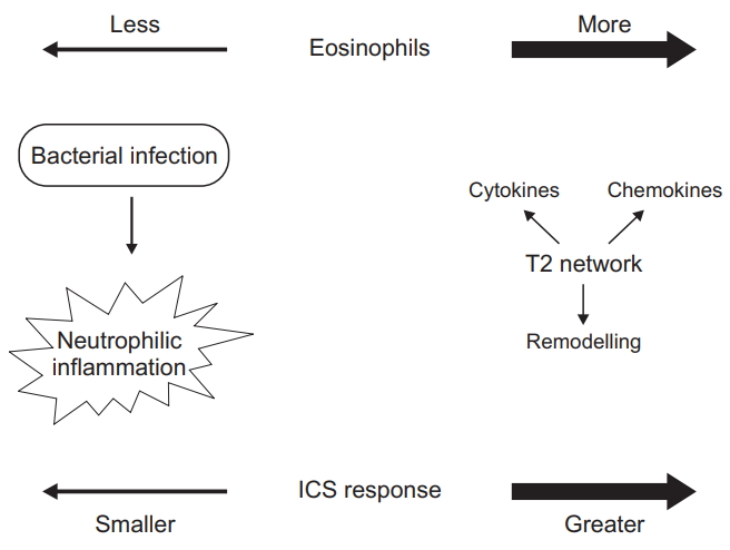
The presence of bacterial infection promotes neutrophilic airway inflammation [56,62,63]. ICS have limited effects on neutrophilic airway inflammation in humans [64-66]. It therefore appears that COPD patients with low eosinophil counts are more likely to skew towards bacterial infection and neutrophilic inflammation that responds poorly to ICS treatment. In contrast, high eosinophil counts, associated with increased T2 gene expression and a lower burden of bacterial infection, is a profile of airway inflammation that is more ICS sensitive; this model to explain the relationships between eosinophils, bacteria and ICS response are shown in Figure 3.
It is relevant to consider whether ICS are targeting eosinophils themselves, and/or other aspects of inflammation associated with higher eosinophil counts. An RCT using sputum eosinophils to predict ICS effects showed improvements in FEV1 in COPD patients with higher eosinophil counts, but no reduction in eosinophil counts [25]. In contrast, ICS/LABA reduced sputum eosinophil counts compared to placebo, in addition to suppression of lymphocytes and mast cells numbers in the bronchial mucosa [65]. There was no reduction in sputum neutrophil numbers. Another bronchoscopy study in COPD patients also showed an effect of ICS treatment on submucosal lymphocytes, but no effect on neutrophils and eosinophils [66]. These studies have provided consistent evidence that ICS reduce airway lymphocyte numbers and have no effect on neutrophil counts, but the results for eosinophils have been mixed. This mixed evidence can be interpreted as showing that ICS can reduce airway eosinophil numbers, but that this finding is not consistent in all patients. This may be due to low baseline eosinophil numbers in some patients. Alternatively, the therapeutic benefit of ICS in eosinophilic COPD patients may be due to pharmacological effects on inflammation components (T2 inflammation) beyond the eosinophil cell.
RCTs investigating the effects of monoclonal antibodies targeting IL-5, conducted in COPD patients with a history of exacerbations, have reported both positive and negative results for the effect on exacerbation rate reduction [67,68]. A prespecified combined analyses of the GALATHEA and TERRANOVA studies of benralizumab (which targets IL-5 receptor alpha) demonstrated that the greatest treatment benefit was observed in the subgroup receiving triple therapy with blood eosinophil counts âĽ220 cells/ÂľL and âĽ3 exacerbations in the prior year [69]. These findings highlight that significant eosinophilic inflammation, which may respond to monoclonal antibody targeting, can persist despite ICS treatment.
Studies focused on COPD exacerbations have found that blood and sputum eosinophil counts are increased in a subset of patients during exacerbations [70]. Interestingly, higher blood eosinophil counts in the stable state are associated with an increased probability of exacerbations with increased sputum eosinophil numbers [71]. This association between eosinophils in the stable and exacerbation states suggests that ICS treatment probably suppresses exacerbation subtypes involving increased eosinophilic inflammation.
GOLD 2019 and Blood Eosinophils
The evidence already reviewed formed the basis of the GOLD 2019 recommendations to use blood eosinophil counts as a biomarker to help direct ICS treatment in COPD patients with a history of exacerbations [3]. These recommendations combine clinical information (exacerbation risk) with biological data (blood eosinophils) as a precision medicine approach in order to optimize the potential for benefit over risk.
The FLAME and IMPACT studies reported different results for the comparison of the effects of ICS/LABA versus LABA/LAMA combinations on exacerbation rates [8,32]. A major reason for these divergent results was the different exacerbations risks of the study populations [34]. Accordingly, GOLD states that the benefits of ICS are likely to be greater in high exacerbation risk patients with a history of âĽ2 moderate exacerbations and/or âĽ1 severe exacerbation in the previous year compared to patients with 1 moderate exacerbation [3]. Other clinical factors relevant to the use of ICS include a history of asthma, which favors ICS use, and risk factors for ICS side effects, such as repeated pneumonia, which argue against ICS use [72]. All of this clinical information should be collected and then used alongside blood eosinophil counts in order to make optimal decisions for each individual.
GOLD uses thresholds of >300 eosinophils/ÂľL and <100 eosinophils/ÂľL to identify individuals with a higher and lower probability, respectively, of experiencing treatment benefit with ICS [46]. The GOLD 2020 revision states that these are âestimates, rather than precise cut-off valuesâ [46]. The purpose of this statement is highlight that small numerical changes in blood eosinophil counts should not lead to a change in ICS treatment, even if the change leads to movement across a threshold value.
Some clinicians have focused on the >300 eosinophils/ÂľL threshold stated in GOLD, preferring to use ICS only in these individuals. However, this approach does not account for the possibility of a treatment benefit that has been observed in patients with 100-300 eosinophils/ÂľL [3,30]. GOLD does not include recommendations concerning eosinophils and current smoking, but there is now evidence that current smoking reduces corticosteroid sensitivity [27,30]. This means that ICS benefits are less likely in current smokers, and that greater ICS benefits may be observed at lower blood eosinophil counts in ex-smokers (i.e., <100 eosinophils/ÂľL).
Conclusion
Blood eosinophil counts are being increasingly used in clinical practice to support clinical decision making regarding ICS use. GOLD recommendations focus on integrating clinical information with eosinophil counts to optimize the choice of combination inhaler to be used for exacerbation prevention [3]. Eosinophilic COPD patients appear to have a wider profile of T2 inflammation, which may provide the mechanistic explanation for the increasing benefit of ICS at higher blood eosinophil counts. Emerging data indicate that lower blood eosinophil counts are associated with increased risk of chronic bacterial infection. Complex relationships appear to exist between eosinophil counts, ICS response, and the airway microbiome. Understanding this complexity is the key to optimizing current pharmacological management and developing novel drugs for the future.
 PDF Links
 PubReader
 ePub Link
 Full text via DOI
 Print
Print Download Citation


