


Introduction
The prevention and management of exacerbations are main objectives of chronic obstructive pulmonary disease (COPD) treatment. Each new exacerbation is harmful for the patient for diverse reasons: it increases in itself the risk of future exacerbations [1], deteriorates the quality of life, accelerates the deterioration of lung function and increases the risk of hospitalization and death [2]. Its prevention is, therefore, a central aspect of the management of these patients. There are various pharmacological and non-pharmacological strategies aimed at both the control and prevention of COPD exacerbations. Although airway inflammation is one of the significant contributors to symptoms and exacerbations, current COPD guidelines do not consider the evaluation of the type of bronchitis or other complex pathophysiological processes involved in its genesis. That leads to generalized management strategies, which are often suboptimal. Although “endotyping” is recommended for “individualized” care of COPD exacerbations, this is not often practiced [3].
We present the following three cases to illustrate the limitations of current guidelines and common clinical practice in most outpatient clinics across the world.
(1) A 67-year-old male with a past smoking history of 21 years, moderate airflow obstruction (forced expiratory volume in 1 second [FEV1] of 61% predicted), and recurrent exacerbations (two in the last 12 months): He is on fluticasone/salmeterol 1,000 μg/100 μg daily and tiotropium 18 mcg daily. After his first exacerbation, his FEV1 decreased to 44% predicted and subsequently worsened to 33% predicted after the second exacerbation. Current guidelines would suggest that both exacerbations “be treated with more bronchodilators,” and perhaps with a “short burst of prednisone” and a “broad-spectrum antibiotic” [4], and perhaps adding long-term macrolide or a phosphodiesterase 4 inhibitor [4,5].
(2) A 57-year-old male, current smoker with a history of 15 pack-years: He reports productive cough, and in increase in wheeze and exertional dyspnea. His FEV1/forced vital capacity (FVC) is 2.8 L/4.4 L (ratio of 63%) and improves to 2.9 L/4.2L post bronchodilator, which is consistent with mild to moderate airflow obstruction (FEV1 of 78% predicted). Chest X-ray is normal. His current treatment includes salbutamol as needed, which he uses about 2 to 4 times a day. Current guidelines would suggest that he be commenced on a combination of a long-acting beta-2 agonist (with or without a long-acting anticholinergic inhaler) [4].
(3) An 81-year-old male, with a 34 years history of smoking: His previous medical history includes glaucoma, benign prostate hyperplasia, diabetes and coronary artery disease. He presents with exertional breathlessness and cough and has had two exacerbations within the last year. His pre-bronchodilator FEV1/FVC is 0.9 L/4.4 L, and postbronchodilator is 1.0 L/4.5 L, which are 29% and 90% predicted, respectively. Total lung capacity is 122%, residual volume is 160%, and KCO is 30% predicted. Arterial blood gases show a PCO2 of 58 mm Hg, PO2 of 64 mm Hg and pH of 7.38. Right ventricular systolic pressure is 40 mm Hg. Computed tomography of the thorax reveals heterogenous centrilobular emphysema. Current treatment is budesonide/formoterol (200 μg/6 μg) 2 puffs twice daily, terbutaline as needed, furosemide and ramipril. Current guidelines would suggest adding a long-acting anticholinergic inhaler or alternatively switching to a single combination inhaler [4].
Current COPD Guidelines on Treatment and Prevention of Acute Exacerbations
Current recommendations are largely focused on decreasing exacerbations and improving symptoms by optimizing the use of bronchodilators. It is known that both long-acting beta agonists (LABA) and long-acting anti-cholinergics (LAAC) can reduce the rate of exacerbations in patients with COPD. Furthermore, the current literature supports that combined therapy (LABA/LAAC) is superior to monotherapy [6] and to LABA/inhaled corticosteroid (ICS) [7] combination, although its effect does not reach the sum of both [8]. More recently, attempts have been made to demonstrate that triple inhalation therapy is even more effective than the LABA/LAAC combination; but the results have been variable. A large study of more than 10,000 patients, which compared triple therapy against two types of dual therapy (LAAC/LABA and LABA/ICS), showed lower rates of moderate and severe exacerbations in the first group [9]. A similar effect is observed when comparing singleinhaler triple combination with single-inhaler dual bronchodilator combination, with a 15% reduction in the risk of moderate to severe exacerbations [10]. The latter was not reflected in a subsequent trial performed in a “real-world clinical setting,” where there were no differences between the groups treated with triple therapy and LAAC/LABA. However, when analyzing by subgroups, it was observed that among patients with blood eosinophilia >6% and in those with two or more previous exacerbations, there was a statistically significant benefit (hazard ratio [HR] of 0.66 and 0.83, respectively) [11]. Therefore, based on the current evidence, there is an inclination to add ICS to dual bronchodilator therapy in patients with moderate to very-severe stable COPD with a high risk of exacerbations, especially in those with serum eosinophils higher than 300 cells/µL [4,5]. A summary of the current principal studies which compare dual and triple inhalers’ effect on FEV1 and exacerbation rates is shown in Table 1.
Despite the availability of these pharmacologic interventions, about 30% of patients with COPD have frequent exacerbations [1], which entails high healthcare costs [5]. As part of the numerous efforts aimed at trying to reduce the rate of hospitalizations in these patients, comprehensive care management programs (CCMP) have been implemented, including strategies such as patient education, implementation of action plans, and serial telephone evaluations by a trained team. Unfortunately, the application of these strategies has not been consistently shown a reduction in the risk of hospitalization, but rather, in some cases they have increased it. This was shown in a trial performed in 426 COPD patients who were assigned to a CCMP versus conventional care. The study was terminated before enrolment was completed, given higher risks of hospitalization and mortality in the intervention group (HR of 1.13 and 3.0, respectively) [12]. Furthermore, there was no improvement in quality of life outcomes with these interventions [13]. These results contrast with other studies in which these strategies were proven effective to reduce COPD-related admissions, although the studied populations were significantly different (e.g., patients with other comorbidities were excluded) [14]. Another interesting explanation for this phenom ena is that patients, having the resource of at-distance health monitoring, experience a false sense of safety, which delays consultation in case of an emergency [15].
Sputum, COPD, and Exacerbation Phenotype
Classically, an acute exacerbation of COPD (AECOPD) is defined as respiratory symptoms that worsen beyond the normal day-to-day variability requiring additional therapy [4,5]. However, this definition of an AECOPD may be overlapping with many concurrent pathologies such as worsening left or right heart function, respiratory or metabolic acidosis, with or without bronchitis [15]. The pillars of AECOPD management according to most guidelines include bronchodilation, systemic corticosteroids, and antibiotics [4], without in depth characterization of the bronchitic component. The non-specific manner of AECOPD treatment with the above strategy does not adequately target the key pathology involved while subjecting the patient to additional side effects. A potential resolution to overcome this challenge is to assess the sputum characteristics at baseline and at each exacerbation for COPD patients and to guide treatment based on these objective measures.
Sputum cytology has already been well established, particularly in asthma [16,17]. In brief, it involves sputum being induced from the lower respiratory tract using increasing concentrations of nebulized saline. Total cell count (TCC) and viability is assessed following processing, and a cytospin slide is made for differential cell count. This protocol has been validated with well-established normal limits described in literature (Table 2) [18-20]. Apprehension regarding the bronchoconstrictive properties of nebulized saline is unfounded as several studies have demonstrated the safety of sputum induction in both stable and exacerbating COPD [21,22]. If the FEV1 is too low for hypertonic saline induction, sputum can be obtained with a modified protocol using normal saline [23]. Spontaneously expectorated sputum is comparable to induced sputum in terms of cell differential and can be used for clinical purposes [24]. When assessed during an exacerbation, the sputum cytology can allow a more discriminatory approach in therapy [25].
Using sputum analysis, it is possible to characterize the patient’s airway inflammation at baseline and at each exacerbation, as there is good evidence that they may be discordant. A comparison of airway characteristics of COPD patients during stability and exacerbation were studied in a retrospective cross-sectional survey and showed that neutrophilic bronchitis is much more prominent during exacerbations compared to baseline [26]. In a similar study that included a subpopulation (n=65) of COPD patients who had successive sputum analysis during convalescence and exacerbations showed that there was poor correlation between the baseline and exacerbation airway cytology. Furthermore, 85.2% of patients had subsequent exacerbations that differed in bronchitic subtype from baseline or even a previous exacerbation [30]. Taken together, this suggests that exacerbations are not simply a worsening of the underlying inflammation and emphasizes the importance of sputum analysis at every exacerbation to tease out these variations that can lead to change in therapeutic strategy for each episode. The approach of combined antibiotics and corticosteroids is only appropriate in 2.5%-8% of exacerbations as defined by the presence of mixed granulocytic bronchitis. Hence, by characterizing the luminal inflammation using sputum examination, the most appropriate and effective therapeutic strategy can be put in place and reduce the likelihood of adverse drug events and economic burden for the patient and the health care system, respectively [31,32].
Eosinophils in COPD: Actor or Spectator?
A proportion of COPD patients have evidence of eosinophilic bronchitis either during exacerbation or at baseline. Sputum eosinophilia is found in 10%-40% of patients with COPD [33] and has been associated with severity of the disease, exacerbation frequency and degree of emphysema on quantitative imaging [34,35]. AECOPD with viral infections, in particular, demonstrate increased eosinophilic activity as evidenced by a significant increase in the presence of soluble eosinophil cationic protein in sputum [36]. The presence of eosinophils in sputum also predicts response to corticosteroids, both systemic [37] and inhaled [38]. In randomized control trials, controlling luminal eosinophils using corticosteroids have been shown to reduce severe AECOPD [25].
Although it is more impractical to obtain, sputum eosinophil counts cannot be replaced by blood eosinophils as a biomarker. In a study investigating the blood eosinophil count that correlates with exacerbations, a threshold of 300 cells/µL or more was found to predict an increased risk of AECOPD [39]. However, when directly compared with sputum, circulatory eosinophil counts correlates poorly with luminal eosinophil counts [34,40]. The role of circulatory eosinophils in airway disease remains questionable as well. When analyzing the effect of blocking eosinophil recruitment from circulation into target tissues, paradoxical elevation of blood eosinophil count was seen in clinical trials using anti-interleukin 13 (IL-13) monoclonal antibodies (MABs) for the treatment of asthma [41]. Despite this elevation, patients in the treatment arm demonstrated improved lung function. One can argue that this lack of correlation or direct pathological role is arbitrary and, if blood eosinophil counts can predict response to therapy, then it remains a valid biomarker. However, this stance may be flawed when looking closer at anti-interleukin 5 (IL-5) drug trials in COPD and comparing them to those done in asthma.
The use of anti-eosinophil therapy targeting the IL-5 pathway in asthma have shown overall improvement in asthmarelated quality of life, reduced corticosteroid use and improvement in lung function. Importantly, it demonstrated dose related reduction in exacerbation frequency [42-45]. The same cannot be said of COPD trials using the same molecules. A study that combined the results of two phase 3 clinical trials where COPD patients with an eosinophilic phenotype were given low and high dose mepolizumab (100 mg in METREX, 100 mg, and 300 mg in METRO). This study concluded that 100 mg mepolizumab is effective at reducing exacerbation frequency among this population. However, it appears that while the lowest dose of mepolizumab appeared to be effective at reducing exacerbation frequency (p=0.04), a three-fold higher dose paradoxically did not (p=0.14) [46] which opposes the data seen in asthma [43]. Moreover, the exclusion of subjects with asthma in this study was based on self-report and may have contaminated the sample. In this study, eosinophilic phenotype was determined by blood eosinophil levels of >150 cells/µL at time of screening or >300 cells/µL during the previous year. These thresholds may not be specific to eosinophilic patients as studies on normal leukocyte differentials done in the 1970s and 1980s among healthy volunteers has established a 95% normal range for blood eosinophils of 0 to 700 cells/µL with a median of 150 cells/µL [47,48]. To resolve these methodological issues, the diagnosis of asthma need to be rigorously excluded and eosinophilia be identified at the tissue level by sputum analysis. When these amendments in patient selection are applied, mepolizumab only depletes sputum and blood eosinophils in COPD without an effect on exacerbation rate [49]. Similarly, benralizumab, an anti-IL-5-receptor MAB, also failed to demonstrate reduction in exacerbation regardless if selecting for patients based on elevated circulatory [50] or sputum [51] eosinophilia. Thus, these negative studies have led to the speculation that eosinophils, whether circulatory or luminal, predict steroid responsiveness and disease severity but is otherwise not directly involved in COPD. Perhaps eosinophilia is only is a marker that rises and falls in concordance with an another, unknown process that is key in COPD pathobiology are not intrinsically contributory themselves.
More recent studies regarding the role of eosinophils in COPD have revealed evidence that challenges the spectator theory. In murine models, lung matrix metalloprotease 12 (MMP-12), a key mediator in emphysematous alveolar destruction produced by macrophages, was found to be elevated [52]. The levels of MMP-12 correlated with the presence of activated eosinophils and their subsequent IL-13 release. In these experiments, the eosinophils were preferentially activated in vitro by IL-33, and not IL-5. This finding suggests that, not only do eosinophils contribute to the pathophysiology of COPD, the luminal eosinophils in COPD have different biology compared to those found in asthma and have entirely different downstream effects. Thus, this emphasizes the importance in controlling eosinophilic inflammation in COPD not only during exacerbations but also while stable, as ongoing eosinophilia may continue to propagate tissue damage even if noxious inhalants have been removed.
While these studies regarding the role of eosinophils represent important breakthroughs in biology, the true impact of the research is still yet to be seen in clinical decision making and, in translation, to patient care. When following currently available guidelines, the use of ICS is not recommended as part of first line therapy [4]. The current guideline recommendations regarding ICS addition are based on blood eosinophil counts, as opposed to sputum [5]. As mentioned above, there is an inclination to add ICS in patients with at least moderate COPD but this may be putting some at increased risk of infections [53]. At the same time, as blood and sputum eosinophils do not correlate well, another proportion of patients who would benefit from ICS may be overlooked when following these principles. Among those without severe disease or who are not considered at “high risk” based on spirometry and previous frequency of exacerbation, and not type of exacerbation, the initiation of ICS is only recommended after a minimum of 12 months of persistent symptoms and reduced health status despite LABA and LAAC dual therapy [5]. However, up to 18% of COPD patients have eosinophilic bronchitis at baseline [26] and may not necessarily fall under the “high risk” profile. Given the new evidence of the role of eosinophils in COPD, rigorous adherence of guidelines without sputum analysis may result in both over- and under-treatment of a large number of patients.
Beyond Cells: Other Uses for Sputum
The utility of sputum is not limited to just cytology but can also be used to assess for other comorbid conditions that may contribute to a patient’s symptoms. The detection of lipids in sputum macrophages can be used as a non-invasive marker for gastroesophageal reflux. Using a reproducible method of indexing the degree of lipid staining using oil red O in macrophages, an index of seven or more correlated with the gold standard 24-hour ambulatory esophageal pH recording, and was highly specific and sensitive [54].
Hemosiderin-laden macrophages (HLM) can also be detected on induced sputum as a maker of pulmonary capillary leakage from left ventricular dysfunction. This has been previously well described on bronchoalveolar lavage (BAL) fluid [55,56] but acquisition of BAL fluid is an inefficient way of establishing left ventricular dysfunction. In a prospective cross-sectional pilot study, 46 dyspneic patients and nine healthy controls underwent echocardiography and sputum induction within 72 hours of each other. The presence of HLM proportion directly correlated with decrease cardiac function and was found to be a sensitive and specific maker for left ventricular dysfunction [57]. This was further validated among patients who were presenting with acute respiratory symptoms as well where HLM were significantly elevated in those whose dyspnea was of cardiac as opposed to pulmonary origin [58]. Persistent elevations of sputum HLM may also increase the likelihood of infectious AECOPD, which is further explored in the next section.
Apart from using sputum for immediate patient management decisions, other non-cellular components can be quantified to better understand the biology of airway diseases. The fluid phase of sputum has several utilities including the measurement of soluble cellular by-products [16]. Such measurements include downstream secretion products of various leukocytes, as a surrogate marker of activity, as well as various cytokines, chemokines, and adhesion molecules that are key in supporting local inflammation [16,59,60]. More recently, autoantibodies have been detected in sputum, which may provide further insight on the contribution of local immunity to underlying pulmonary conditions [61,62]. While systemic immunoglobulin deficiency is a contributor to infectious exacerbations, as discussed below, local immunoglobulin levels also appear to impact exacerbation frequency and lung function in COPD [63]. Bacterial migration across the epithelium in small airways is poorly inhibited where there is local deficiency of secretory IgA. This promotes further inflammation and remodeling, even with smoking cessation [63]. Thus far, these assessments have been limited to research use but, may one day be employed clinically to further fine tune management strategies and personalized medicine.
What Causes Infective Exacerbations?
The most common causes of COPD exacerbations are lower respiratory tract infections [64], although other factors such as environmental pollution and exposure to fine particulate matter may also contribute [65]. Whilst viruses are the most frequent causative microbe, bacterial colonization also plays a significant role in infective exacerbations [66].
Why some individuals with COPD are more susceptible than others to develop infective exacerbations is still an unresolved question. Though some explanations for this propensity are already well established, such as structural alterations and immune dysregulations secondary to smoke [63,67], there are still novel mechanism which remain poorly understood. Several biomarkers have been linked to the predisposition to respiratory infections in these patients, including quantification of multiple proteins, specific cells, metabolites in exhaled air, images and even genetic predictors [15,68]. Recurrent respiratory tract infection may also occur because of local humoral deficiency. We recently measured a complete immunoglobulin profile in asthmatics with recurrent respiratory tract infection and found that there was a relative deficiency of sputum IgA compared to healthy controls [69].
It has been observed that the development of these infections could be correlated with changes in the composition of the pulmonary microbiome through increased inflammation of the airways [70]. A study that followed 87 subjects and analyzed their pulmonary microbiota with molecular biology techniques showed that both the diversity and the proportion of different families of bacteria varied within the same individual throughout periods of stability versus exacerbation. Interestingly, in acute respiratory episodes, individuals tended to carry more abundantly Proteobacteria (Moraxella catarrhalis and Haemophilus influenzae) than Firmicutes (Streptococcus pneumoniae). This relationship was reversed during periods of stability. Likewise, it was observed that the treatment with oral corticosteroids was related to lower diversity and a change in the microbiome composition of the investigated subjects. These alterations were maintained even six weeks after the episodes were treated [71].
Among the genetic factors involved with an increased risk of respiratory infections, those related to iron metabolism stand out [72]. As an essential nutrient, iron plays a central role in bacterial survival and multiplication. Furthermore, in states of chronic inflammation, hepcidin-mediated intracellular iron overload could potentially cause immune cell dysfunction [73,74] Locally, the iron overload at the respiratory system can be measured by staining hemosiderin in sputum macrophages [57], which can undoubtedly be an interesting biomarker that could predict higher infectious risk in these patients. A retrospective study showed a correlation between a higher percentage of HLM (hemosiderin index) and a higher frequency of infectious exacerbations in the previous two years. Interestingly, it was also shown that higher local levels of interleukin 6, the cytokine responsible for promoting hepcidin release, correlated with a higher hemosiderin index [75]. Excess iron in pulmonary macrophages also appears to be a predisposing factor for infective exacerbation when studied prospectively, with those with a higher sputum hemosiderin index having a significantly shorter time to infective exacerbation (unpublished data).
Management of AECOPD: St. Joseph’s Strategy and Resolving Our Cases
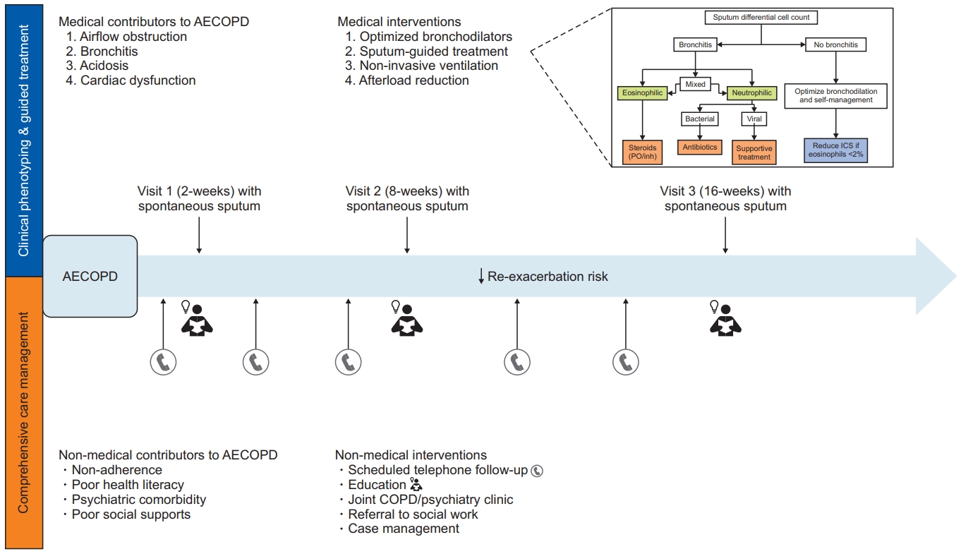
Our local approach to AECOPD could be summarized (Figure 1) as follows.
First, we try to identify the cause of the symptoms, whether it is worsening of airflow limitation, bronchitis, respiratory or metabolic acidosis, left or right ventricular dysfunction, or a combination of these. A general approach to this can be easily made through simple tests available in the emergency department, including spirometry, chest X-ray, and arterial blood gases. Treatment is initiated according to guidelines, with bronchodilators, systemic steroids and antibiotics if they are indicated. Secondly, as soon as feasible to obtain, a sputum sample is collected, and treatment is adjusted accordingly. If sputum shows eosinophilic bronchitis, steroids are continued or increased. Neutrophilic bronchitis (neutrophils ≥64.4% with a TCC greater than 25×106/g cells) suggests an underlying bacterial infection and is thus treated with antibiotics. Notably, even when mixed granulocytic bronchitis (≥2% eosinophils and ≥65% neutrophils) has been associated with poorer clinical outcomes [76], its physiopathology has not been completely understood and thus is managed with both antibiotics and steroids [15]. Normal cell counts would prompt discontinuation of both systemic steroids and antibiotics. If we observe a high hemosiderin index, left ventricle function will be further assessed. Additionally, aspiration would be suspected if there is a high lipid index, and speech and language pathologist evaluation would be requested.
Within 2 weeks of discharge, patients are seen in a specialized post-discharge COPD clinic, where we utilize a similar clinical phenotyping approach in combination with a comprehensive care management program. Patients are seen again at 8- and 16-weeks post-discharge with each visit accompanied by clinical assessment, including complete blood count (for peripheral eosinophilia), blood gases (for persistent hypercapnic respiratory failure), spirometry, and spontaneous sputum cytometry and culture. These measurements allow for the individualized prescription of inhalers, to optimize airflow and suppress persistent inflammation. CCMP is delivered by a trained nurse and includes case management, regular telephone follow-up, inhaler technique assessment and education during clinic visits, and referral to appropriate services (e.g., smoking cessation clinic). Patients can contact the clinic nurse if they develop worsening symptoms or require clarification regarding their treatment. Combining these two seemingly disparate interventions accounts for the medical (e.g., uncontrolled bronchitis) and non-medical (e.g., poor adherence) contributors to uncontrolled disease. Retrospective study of this combined clinic intervention has demonstrated a significant reduction in healthcare utilization, with sputum cytometry influencing treatment decisions in many cases (unpublished data). A clinical trial of this intervention is planned to rigorously test its efficacy and cost-effectiveness.
1. How would this be applied to the proposed cases?
At his first exacerbation, his sputum showed 23.4×10⁶/g total cells, with 0.2% eosinophils and 88% neutrophils. He was prescribed an antibiotic course and short-acting beta-agonist in addition to continuing his maintenance inhalers. Symptoms resolved in six days. At his second exacerbation, sputum TCC was 7.3×10⁶/g, with 22% eosinophils and 46% neutrophils, for which he received a course of prednisone. This example outlines how two exacerbations could be related to different underlying mechanisms and therefore, should be treated differently.
According to current guidelines, this patient would be categorized as mild COPD and thus should receive either LABA or LAAC. However, his sputum test showed a TCC of 6.2×10⁶/g, with 57% neutrophils and 11% eosinophils. Though this patient would benefit from ICS, his assigned disease severity would at most prompt the addition of another long-acting bronchodilator, thus leaving his eosinophilic bronchitis untreated. It is relevant to note that unpublished data from our clinical experiences has shown that after bronchitis treatment, whether neutrophilic or eosinophilic, a percentage of previous “COPD” patients will no longer meet the spirometric diagnostic criteria, suggesting that unresolved bronchitis may lead to overdiagnosis.
This patient’s investigations showed a sputum TCC of 64.8×10⁶/g cells with 82% neutrophils, and 9% eosinophils. His sputum hemosiderin index was 13%. His bloodwork revealed a chloride of 84 mEq/L, and potassium of 3.1 mEq/L. In this case, given severe COPD with a high risk of exacerbations and uncontrolled eosinophilic bronchitis, chronic treatment should be adjusted to higher doses of ICS, and low prednisone maintenance dose and long-term macrolides should be considered. Ventricle offload treatment should be implemented if cardiac involvement is demonstrated by clinical exam and echocardiogram, and electrolytic disturbances must be corrected, including spironolactone, chloride, and potassium replacement. Acetazolamide, which is often prescribed, should only be added if the patient has enough respiratory muscle reserve to manage higher respiratory rates. Finally, BiPAP support could be incorporated if it is appropriate to do so.
2. Key points
- COPD exacerbations are common even in patients treated according to current guidelines. These events are associated with worsening in lung function, worse symptom control, higher mortality rates and an elevated economic burden.
- Current treatments aimed to reduce AECOPD rates include both pharmacologic and non-pharmacologic approaches. Some strategies, like CCMP, have failed to improve health indicators in these patients, and actually, have been associated with higher hospitalization and mortality risk.
- Sputum assessment, while stable and during exacerbation, is essential for phenotyping and for guidance of therapeutic strategies. It is a safe and rapid method of evaluation and will likely broaden beyond cytology and provide insights regarding other causes of respiratory symptoms.
- The main cause of COPD exacerbations are acute respiratory infections. There are several alterations which underlie higher susceptibility to them, including structural changes, immune impairment, and novel mechanisms that are still poorly understood. Some of them, like iron overload, have been investigated and could be considerate as target checkpoints for future treatment strategies.
- Our local approach to AECOPD include the assessment of the bronchitic component and is focused on airway inflammometry. This personalized strategy has been proven effective to decrease exacerbations rates.
 PDF Links
 PubReader
 ePub Link
 Full text via DOI
 Print
Print Download Citation


