Effects of Antioxidant on Oxidative Stress and Autophagy in Bronchial Epithelial Cells Exposed to Particulate Matter and Cigarette Smoke Extract
Article information

Abstract
Background
We evaluated the effect of particulate matter (PM) and cigarette smoke extract (CSE) on bronchial epithelial cell survival, as well as oxidative stress and autophagy levels. Moreover, we aimed to assess the effect of the antioxidant N-acetylcysteine (NAC) on the adverse effects of PM and CSE exposure.
Methods
Normal human bronchial epithelial cells (BEAS-2B cells) were exposed to urban PM with or without CSE, after which cytotoxic effects, including oxidative stress and autophagy levels, were measured. After identifying the toxic effects of urban PM and CSE exposure, the effects of NAC treatment on cell damage were evaluated.
Results
Urban PM significantly decreased cell viability in a concentration-dependent manner, which was further aggravated by simultaneous treatment with CSE. Notably, pretreatment with NAC at 10 mM for 1 hour reversed the cytotoxic effects of PM and CSE co-exposure. Treatment with 1, 5, and 10 mM NAC was shown to decrease reactive oxygen species levels induced by exposure to both PM and CSE. Additionally, the autophagy response assessed via LC3B expression was increased by PM and CSE exposure, and this also attenuated by NAC treatment.
Conclusion
The toxic effects of PM and CSE co-exposure on human bronchial epithelial cells, including decreased cell viability and increased oxidative stress and autophagy levels, could be partly prevented by NAC treatment.
Introduction
Air pollution is associated with respiratory and cardiovascular diseases. Particulate matter (PM), nitric oxide, and ozone are representative air pollutants associated with the deterioration of the respiratory system [1,2]. PM is a complex mixture of solid particles and liquid droplets in air that have a diameter of less than 10 µm. Notably, PM with aerodynamic diameters of ≤2.5 µm (PM2.5) may reach deeper into the small airway and alveolar space and exert more direct toxicity than PM10 [3].
Chronic obstructive pulmonary disease (COPD), characterized by persistent and progressive airflow obstruction, is a leading cause of morbidity and mortality worldwide [4]. Several epidemiologic studies have highlighted the relationship between PM and increased risk of exacerbation, hospital visits, and mortality in COPD patients [2,5-7]. Considering the prevalence and health-associated effects of COPD in Korea [8,9], the increase in PM levels is expected to have a negative health impact on COPD patients, highlighting the need to address this issue.
Although PM is composed of diverse chemicals and induces inflammation and oxidative stress in various cells [10], the mechanisms underlying PM-induced cytotoxicity are not completely understood. However, oxidative stress, mitochondrial free radical formation, and activation of pro-inflammatory factors are thought to be involved in this process [11]. Oxidative stress caused by PM exposure may induce reactive oxygen species (ROS) generation and is regarded as a pivotal mechanism of cytotoxicity. ROS, such as hydrogen peroxide (H2O2), may also be generated by cigarette smoke. The increase in ROS by various stimuli is known to be associated with the increased autophagy levels [12].
Autophagy is a self-digestion process for the recycling of cellular organelles and cytoplasm in cells and is activated by various stimuli. During the autophagy process, intracellular materials are sequestered into vesicles of the membrane bilayer to form autophagosomes, a process which is mediated by autophagy-related proteins, such as Atg5, Atg12, Beclin1, phosphatidylinositide 3 kinase, and microtubule-associated protein 1 light change 3 (LC3) [13].
The respiratory tract is the primary site of exposure to air pollutants and cigarette smoke in humans, both of which can damage normal cells and are known to contribute to COPD development. Here, we focused on the additive cytotoxicity induced by PM and cigarette smoke extract (CSE) and aimed to investigate whether PM-induced adverse effects could be intensified by additional exposure to cigarette smoke and if treatment with an antioxidant could restore this effect in bronchial epithelial cells in vitro .
Materials and Methods
1. Cell culture and drug
Normal human bronchial epithelial BEAS-2B cells were cultured in BEpiC medium (ScienCell Research Laboratories, Carlsbad, CA, USA) containing bronchial epithelial cell growth supplement and penicillin/streptomycin at 37°C in a 5% CO2 incubator. Cells between passages 3 and 10 were used in all experiments. N-acetyl-L-cysteine (NAC; Sigma-Aldrich, St. Louis, MO, USA) was used to evaluate the effects of antioxidant on cytotoxicity. BEAS-2B cells were grown in 6-well plates overnight and cells at 70%–80% confluence were pre-treated with 10 mM NAC or phosphate-buffered saline (PBS) for 1 hour before treatment with PM and CSE.
2. Urban PM
Urban PM was obtained from the National Institute of Standards and Technology (SRM 1648a). The SRM1648a (PM10) had a mean particle diameter of 5.85 μm and was dispersed in solution via ultrasonication in PBS for 10 minutes at a concentration of 10 μg/μL. BEAS-2B cells were seeded in 6-well plates overnight and then treated with urban PM with or without simultaneous CSE exposure. PM at concentrations of 0, 25, 50, 100, and 200 μg/cm2 were treated for evaluation of cell viability, and in the experiments on RNA or protein expression, PM were treated at the concentration at which cell viability was 50%.
3. Preparation of cigarette smoke extract
Commercialized cigarettes (THIS; 84-mm length with 8-mm diameter; sold by KT&G, Seoul, Korea) were continuously smoked using a tubing silicone system connected to a vacuum system. Smoke from one cigarette was passed through 10 mL of PBS to prepare the 100% CSE [14]. This CSE solution was then diluted with PBS to prepare a 1% and 5% CSE with and without urban PM.
4. Cell viability assay
Cell viability was measured using the EZ-cytox cell viability/cytotoxicity kit (DoGen Bio Co., Ltd., Seoul, Korea). Briefly, 5,000 cells/well of BEAS-2B cells were seeded in 96-well plates and incubated for 24 hours, followed by replacement of the culture medium. Urban PM was added to cells with and without CSE for 12–24 hours. At the indicated time, the EZ-cytox solution was added to the cells according to the manufacturer’s instructions. Finally, the absorbance of the medium was measured at 450 nm and the viability percentage was calculated.
5. Intracellular ROS measurement
Cellular ROS levels were determined using the DCFDA cellular ROS detection assay kit (Abcam, Cambridge, MA, USA) according to the manufacturer’s protocol. At the indicated time, the cells were stained with 25 μM DCFDA in 1× buffer for 45 minutes at 37°C and gently washed with PBS. The fluorescence intensity was measured at Ex/Em=485/535 nm using a fluorescence plate reader (BioTek, Winooski, VT, USA).
6. Real-time polymerase chain reaction
Total RNA was isolated from the cells using the TRIzol reagent (Invitrogen, Grand Island, NY, USA) and reverse-transcribed. Real-time polymerase chain reaction (PCR) was performed using a CFX96 Real-Time PCR Detection System (Bio-Rad Laboratories, Hercules, CA, USA). The reactions were performed using the following selective primers: catalase (CAT), forward 5’-ACCCTCGTGGGTTTGCAGTGA-3’ and reverse 5’-CGAGCACGGTAGGGACAGTTCA-3’; superoxide dismutase 2 (SOD2), forward 5’-GCAGCTGCACCACAGCAAGC-3’ and reverse 5’-CGTGCTCCCACACATCAATCCCC-3’; glutathione peroxidase 1 (GPX1), forward 5’-TGGGCATCAGGAGAACGCCA-3’ and reverse 5’-GGGGTCGGTCATAAGCGCGG-3’; GPX3, forward 5’-TGACGGGCCAGTACATTGA-3’ and reverse 5’-TCCACCTGGTCGGACATACT-3; glutathione reductase (GSR), forward 5’-AGGAGCTGGAGAACGCTGGC-3’ and reverse 5’-CAATGGCCCAGAGCAGGCA-3’; and glyceraldehyde-3-phosphate dehydrogenase (GAPDH), forward 5’-GTCAGTGGTGGACCTGACCT-3’ and reverse 5’-AGGGGTCTACATGGCAACTG-3’. For amplification, the iQ SYBR gene expression assay (Bio-Rad Laboratories) was used according to the manufacturer’s instructions.
7. Western blot analysis
The cells were extracted in RIPA buffer and centrifuged at 12,000 ×g for 10 minutes at 4°C and the protein content in the supernatants was quantified via BCA (Thermo Scientific, Rockford, IL, USA). The cell extracts were then separated via 12% sodium dodecyl sulfate polyacrylamide gel electrophoresis, transferred onto PVDF membranes (Millipore, Bedford, MA, USA), and blocked with 5% skim milk for 1 hour at room temperature. The membranes were blotted with monoclonal antibodies against specific proteins, as indicated. Antibodies against LC3B and β-actin were purchased from Abcam. The relative intensities of the bands were quantified using the ImageJ software (National Institutes of Health, Bethesda, MD, USA).
8. Statistical analysis
Data are expressed as mean±standard deviation of three independent experiments and statistically analyzed using one-way ANOVA with the Tukey post-hoc test or two-way ANOVA with the Bonferroni test, using the GraphPad Prism statistical software (San Diego, CA, USA). Statistical significance was set at p<0.05.
Results
1. Urban PM and CSE decreased cell viability and NAC pretreatment exhibited protective effects on cell survival
To identify the effect of urban PM on BEAS-2B cells, we used an EZ-CYTOX assay and measured cell viability after exposure to various concentrations of PM with and without CSE.
We found that cellular cytotoxicity after treatment with PM increased in a concentration-dependent manner (Figure 1A). We next investigated the effect of CSE on PM-induced cytotoxicity. BEAS-2B cells exposed to 1% CSE were not affected but 5% CSE exposure significantly decreased cell viability. Moreover, CSE exposure further decreased cell viability in PM-exposed cells at both high (5%) and low (1%) concentrations (Figure 1B).
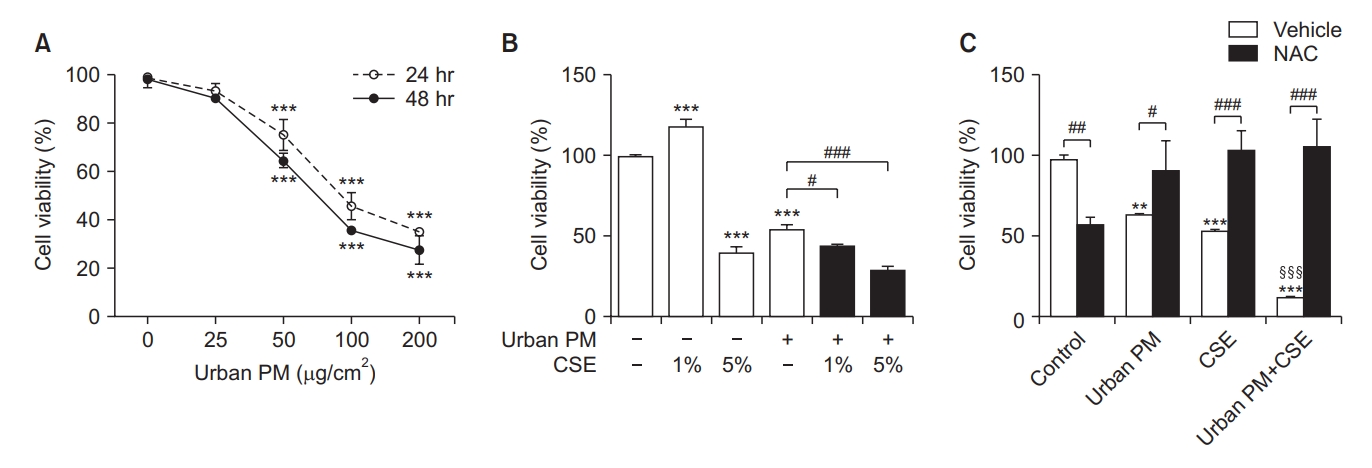
Effect of urban particulate matter (PM) and cigarette smoke extract (CSE) on the viability of BEAS-2B cells. (A) Cell viability after treatment with various concentrations of urban PM for 24 and 48 hours. (B) Cells were treated with 1% and 5% CSE with or without PM for 24 hours. (C) Effect of antioxidants on cell viability in CSE/PM-exposed cells. BEAS-2B cells were pre-treated with N-acetylcysteine (NAC) for 1 hour, then treated with 70 µg/cm2 of PM with or without 5% CSE for 12 hours. Data are expressed as mean±standard deviation (SD). #Compared to vehicle, §Compared with urban PM or CSE, *p<0.05, **p<0.01, ***p<0.001.
To assess the protective effect of the antioxidant NAC against cytotoxicity, we pre-treated BEAS-2B cells with 10 mM NAC for 1 hour. Then, cells were incubated with PM at a concentration of 70 µg/cm2 and 5% CSE for 12 hours. Our results showed that NAC pretreatment reversed the decrease in cell viability caused by exposure to PM or CSE alone. Furthermore, the cytotoxic effects induced by simultaneous exposure to PM and CSE were also restored by NAC pretreatment. This suggests a protective effect of NAC against the cytotoxic effects of PM and CSE exposure (Figure 1C).
2. Urban PM and CSE increased oxidative stress and NAC pretreatment exerted beneficial effects on oxidative stress
Cells were treated with 50 µg/cm2 of urban PM with or without 5% CSE for 24 hours and then the DCFDA assay was performed to quantify ROS generation levels. Treatment with 50 µg/cm2 of urban PM was shown to increase ROS generation when compared to control cells but treatment with 5% CSE alone did not affect oxidative stress levels (Figure 2A).
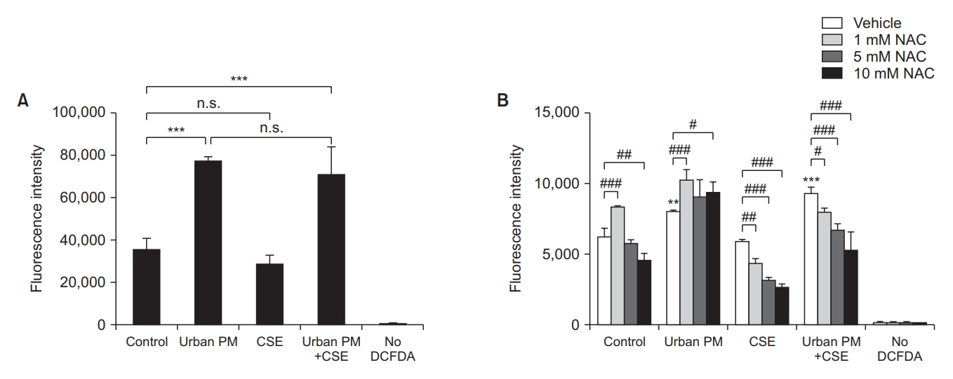
Effects of urban particulate matter (PM) and cigarette smoke extract (CSE) on reactive oxygen species (ROS) generation and oxidative stress. (A) Cells were treated with 50 µg/cm2 of urban PM with or without 5% CSE for 24 hours, then quantitative analysis of DCFDA was performed. (B) Cells were pre-treated with 1, 5, and 10 mM of N-acetylcysteine (NAC) for 1 hour and then incubated with urban PM with or without CSE. ROS generation was detected by quantitative analysis of DCFDA. #Compared to the vehicle, *p<0.05, **p<0.01, ***p<0.001. n.s.: not significant.
Next, to assess the effect of NAC on ROS generation, cells were pre-treated with 1, 5, or 10 mM NAC and then incubated with the same concentration of PM and/or CSE. Afterwards, the DCFDA assay was used to detect cellular ROS levels. NAC pretreatment did not reduce ROS levels in PM-exposed cells, but significantly decreased ROS levels in CSE-exposed cells in a concentration-dependent manner. Notably, the protective effect of NAC against oxidative stress persisted when the cells were simultaneously treated with both PM and CSE (Figure 2B).
3. Urban PM and CSE altered the expression of genes encoding antioxidant enzymes
The relative expression of genes encoding antioxidant enzymes in urban PM and CSE-treated cells after 24 hours is shown in Figure 3. Quantitative reverse transcription polymerase chain reaction (RT-PCR) analysis revealed decreased CAT, SOD2, GPX1, GPX3, and GSR gene expression levels in BEAS-2B cells exposed to 70 µg/cm2 of PM when compared to control cells. Moreover, cells exposed to 5% CSE exhibited decreased CAT, SOD2, and GPX1 gene expression levels but increased GPX3 and GSR gene expression levels when compared to control cells. When cells were simultaneously treated with both PM and CSE, CAT and GPX3 gene expression levels were shown to be decreased, but not significantly when compared to cells exposed to urban PM alone. In the case of SOD2 and GPX1, gene expression levels were decreased in cells exposed to both PM and CSE, but not significantly when compared to cells treated with PM or CSE alone. Only GSR gene expression levels were found to be significantly reduced in cells exposed to both PM and CSE simultaneously when compared to cells exposed to PM or CSE alone.
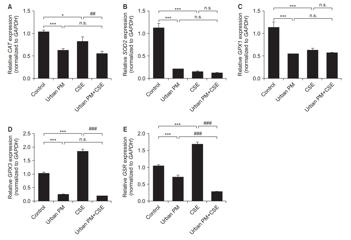
Effect of urban particulate matter (PM) and cigarette smoke extract (CSE) on the mRNA expression of antioxidant-related genes. Cells were treated with urban PM at 70 µg/cm2 and 5% CSE for 24 hours and the expression levels of genes encoding antioxidant enzymes were quantified by real-time polymerase chain reaction, while the mRNA expression levels of catalase (CAT) (A), superoxide dismutase 2 (SOD2) (B), glutathione peroxidase (GPX) (C, D), and glutathione reductase (GSR) (E) were evaluated. GAPDH: glyceraldehyde-3-phosphate dehydrogenase. #Compared to the urban PM or CSE group, *p<0.05, ***p<0.001. n.s.: not significant.
4. Potential protective effect of NAC against urban PM and CSE-induced altered expression of antioxidant enzymes genes
To evaluate the protective effect of NAC against the altered expression of genes encoding antioxidant enzymes, cells were pre-treated with 5 and 10 mM of NAC for 1 hour, then incubated with 50 µg/cm2 of urban PM with or without 5% CSE. RT-PCR was then used to quantify the expression of antioxidant enzyme-encoding genes (Figure 4). Our results showed that the protective effect of NAC pretreatment differed for each gene. For CAT and SOD2, 5 mM NAC pretreatment restored the reduced CAT gene expression induced by PM or CSE exposure alone, while it had no effect on CAT gene expression levels induced by PM and CSE co-exposure. Notably, pretreatment with high NAC concentrations (10 mM) did not exert a beneficial effect. However, reduced GPX1 gene expression levels induced by PM and/or CSE exposure were also recovered by NAC pretreatment, and this protective effect was more prominent at high NAC concentrations. For the GPX3 gene, NAC pretreatment did not exert a protective effect against the PM and/or CSE exposure-induced reduction in GPX3 gene expression levels. Finally, for the GSR gene, pretreatment with 5 mM NAC recovered the decreased expression of GSR induced by CSE exposure but had no effect on the decreased GSR gene expression levels induced by PM or simultaneous PM and CSE exposure.
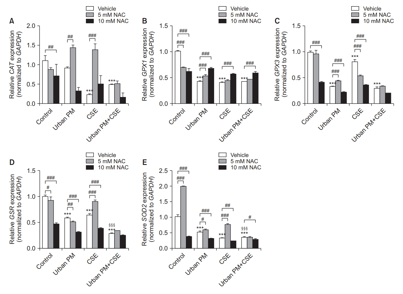
Effect of N-acetylcysteine (NAC) on urban particulate matter (PM) and cigarette smoke extract (CSE) induced altered mRNA expression of antioxidant encoding genes. Cell lysates were used to perform real-time polymerase chain reaction experiments and the mRNA expression levels of catalase (CAT) (A), glutathione peroxidase (GPX) (B, C), glutathione reductase (GSR) (D), and superoxide dismutase 2 (SOD2) (E) were evaluated after 24 hours of exposure to urban PM and CSE. GAPDH: glyceraldehyde-3-phosphate dehydrogenase. #Compared to the vehicle, §Compared with urban PM or CSE, *p<0.05, **p<0.01, ***p<0.001.
5. Urban PM and CSE exposure increased autophagy in BEAS2-B cells
Exposure to urban PM for 24 hours resulted in a dose-dependent increase in LC3I to LC3II conversion (Figure 5A). To investigate the additive effect of CSE exposure in urban PM-exposed cells, experiments were repeated with cells exposed to PM with or without 5% CSE. We found that autophagic activation was increased when cells were exposed to both CSE and PM (Figure 5B).
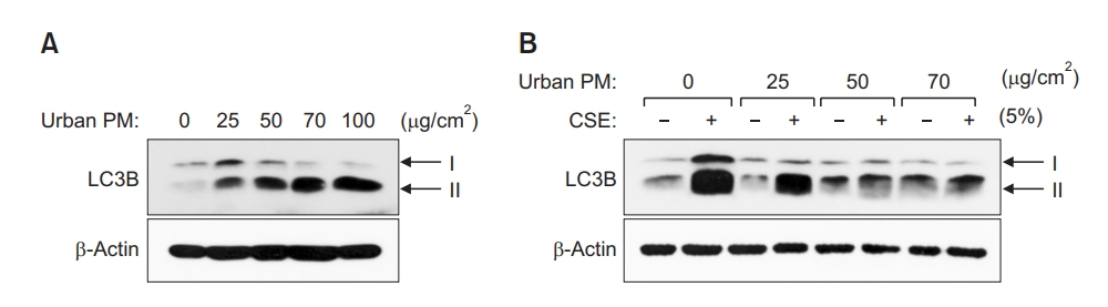
Urban particulate matter (PM) and cigarette smoke extract (CSE) induces autophagy in normal bronchial epithelial cells (HBEpiC). (A) Western blotting of LC3B protein accumulation in HBEpiC. Cell lysates obtained after exposure to urban PM were used for western blot experiments to evaluate protein expression; β-actin was used as the control. (B) Intensified LC3B-II accumulation by CSE co-treatment.
6. NAC pretreatment attenuated autophagy induced by PM and CSE exposure
The increase in LC3B expression caused by urban PM was not affected by NAC pretreatment. However, NAC pretreatment exhibited a profound protective effect against CSE-induced autophagy activation. Moreover, NAC pretreatment also reduced LC3B expression levels induced by exposure to both PM and CSE (Figure 6A).
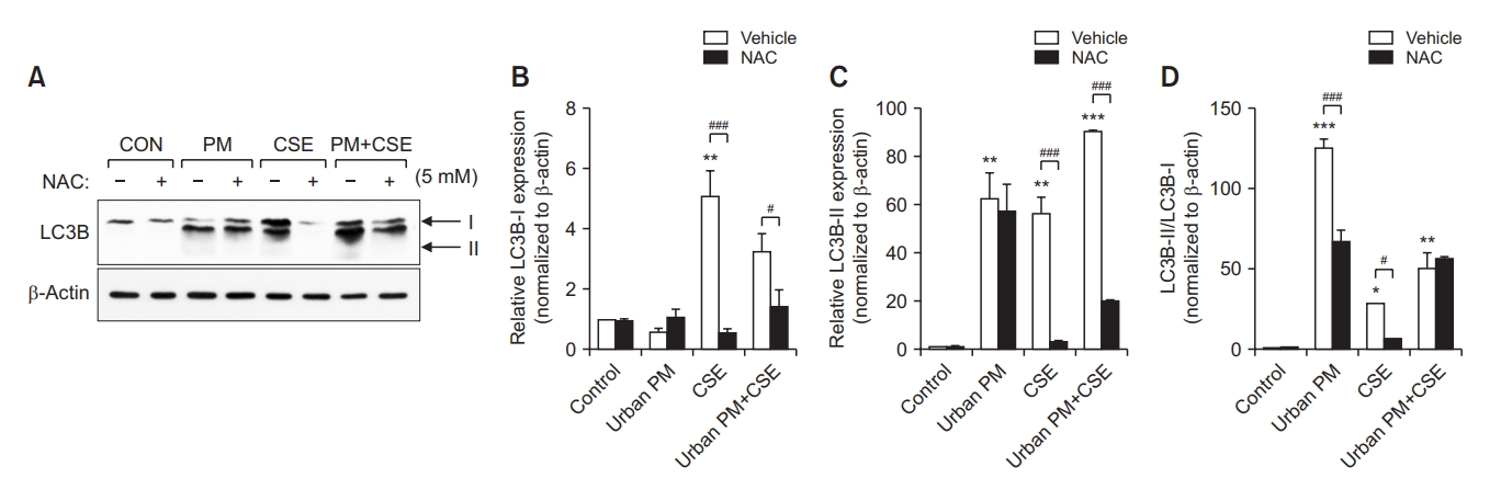
Attenuation of LC3B-II accumulation by N-acetylcysteine (NAC) pretreatment. (A) The LC3B process and accumulation of LC3-II induced by exposure to urban particulate matter (PM) 50 μg/cm2 and 5% cigarette smoke extract (CSE) treatment for 24 hours was attenuated by pretreatment with 5 mM NAC for 1 hour; β-actin was used as the control. Relative LC3B-I (B) and LC3B-II (C) expression, as well as the LC3B-II/LC3B-I ratio (D) were analyzed. #Compared to the vehicle, *p<0.05, **p<0.01, ***p<0.001.
Relative LC3B-I expression levels were markedly increased by CSE exposure and NAC pretreatment significantly reduced this effect (Figure 6B). However, urban PM did not affect LC3B-I expression levels. In contrast, exposure to PM or CSE increased relative LC3B-II expression levels, an effect which was intensified by PM and CSE co-treatment. NAC pretreatment inhibited the effects induced by PM exposure with or without CSE and was shown to significantly increase LC3B-II expression levels (Figure 6C). The ratio between LC3B-II and LC3B-I was markedly increased by exposure to PM or CSE but was reduced by NAC pretreatment. However, in cells exposed to both PM and CSE, the LC3B-II/I ratio did not increase when compared to cells treated with PM alone and NAC pretreatment had no effect on the LC3B-II/I ratio (Figure 6D).
7. Apoptosis and necrosis in urban PM and CSE induced cytotoxicity
To evaluate another mechanism such as apoptosis and necrosis involved in PM and CSE induced cytotoxicity, caspase-3 and cyclophilin A expression were investigated by western blot method. The expression of caspase-3 was increased by CSE and that of cyclophilin A was increased by PM, and NAC pretreatment decreased both (Figure 7).
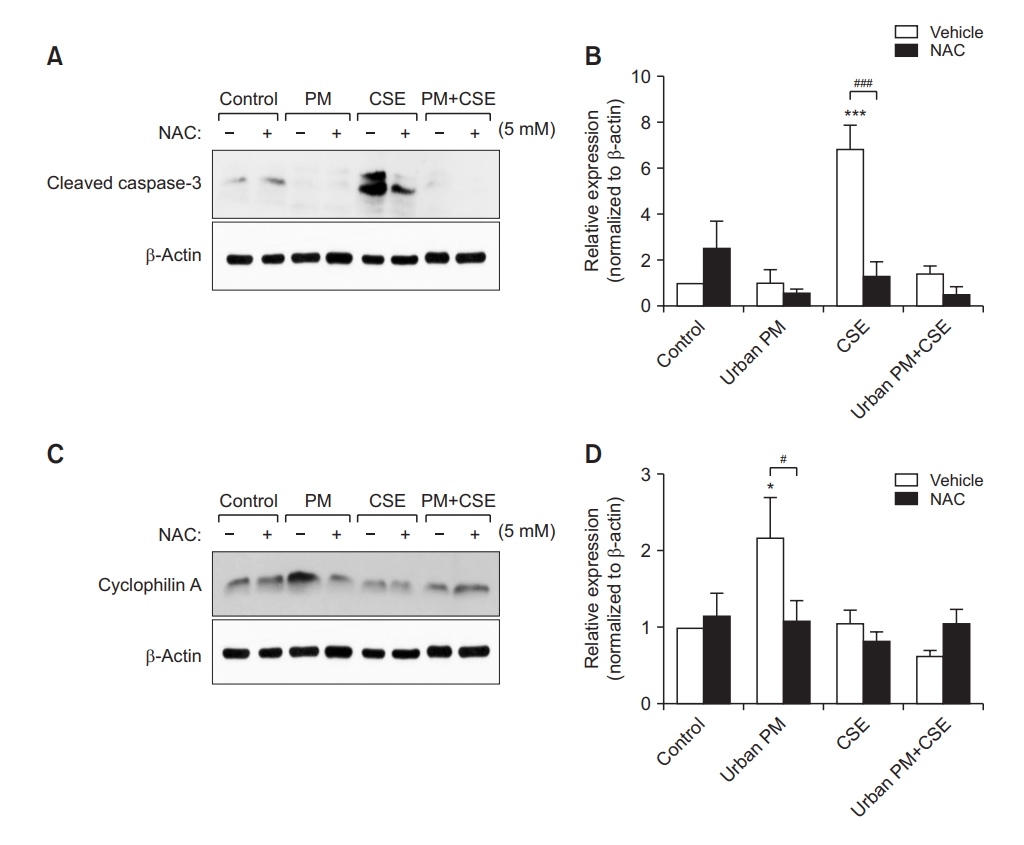
Urban particulate matter (PM) and cigarette smoke extract (CSE) induces apoptosis and necrosis. (A) The cleaved caspase-3, as marker for apoptosis, expression was increased by exposure to 5% CSE for 24 hours, then attenuated by pretreatment with 5 mM N-acetylcysteine (NAC) for 1 hour; β-actin was used as the control. (B) Relative expression of cleaved caspase-3 was presented. (C) The expression of cyclophilin A, as marker for necrosis, was increased by urban PM 50 μg/cm2 for 24 hours, then attenuated by pretreatment with 5 mM NAC for 1 hour. (D) Relative expression of cyclophilin A was presented. #Compared to the vehicle, *p<0.05, ***p<0.001.
Discussion
In the present study, we showed that urban PM exposure alone or in combination with CSE decreased cell survival in normal bronchial epithelial cells, a process that might be mediated by increased oxidative stress and autophagy activations. Moreover, we showed that antioxidant NAC treatment attenuated PM and CSE-induced adverse effects in bronchial epithelial cells.
The pathogenesis of COPD remains unclear and is believed to be induced by multiple factors. Cigarette smoking is considered the most relevant and well-known risk factor of COPD, with only approximately 25%–40% of COPD cases not being associated with smoking [15]. According to a report from the Health Effects Institute in the United States, the annual mean concentration of PM2.5 increased from 26 μg/m3 in 1990 to 29 μg/m3 in 2015 [16]. Likewise, in Korea, the average annual concentration of PM is steadily increasing [17]. Although epidemiologic evidence has confirmed the role of PM exposure in increased COPD exacerbation and mortality [2,5-7], the mechanism underlying this process remains unclear. Furthermore, there is limited evidence for the role of PM exposure in COPD incidence [18,19]. PM is composed of diverse chemicals and induces inflammation and oxidative stress in various cells [10]. Recently, several studies have investigated the effects of PM and CSE on cell damage and identified clues regarding the mechanisms underlying these effects [20-23]. PM2.5 was shown to aggravate inflammatory processes and apoptosis in cigarette-inflamed bronchial epithelium both in vivo and in vitro [20]. This suggested a possible mechanism for the effects of PM2.5 in COPD by showing that cigarette-inflamed airways were more vulnerable to PM2.5 than normal airways. Zhou et al. [21] found that the microRNA miR-194-3p is a biomarker of COPD and is correlated with the PM2.5-induced decline in lung function. The authors also showed that PM2.5 significantly aggravated apoptosis in cigarette-inflamed bronchial epithelial cells and that miR-194-3p was downregulated in PM2.5 and cigarette smoke-treated bronchial epithelial cells [21].
Oxidative stress is an important factor in the pathophysiology of COPD, with H2O2 levels being increased in the airways of patients with COPD [24,25]. Moreover, H2O2 levels were shown to be increased in exacerbated COPD when compared to COPD patients in the stable state [26]. The adverse effects of PM on respiratory diseases have not been clearly elucidated but oxidative stress is considered the most important axis of PM-induced cytotoxicity. In this study, we confirmed that PM exposure exerts adverse effects on oxidative stress in bronchial epithelial cells and concomitant CSE exposure aggravates this cytotoxic effect.
Exposure to both urban PM and CSE increased ROS generation when compared to exposure to each PM or CSE alone. However, exposure to CSE alone did not significantly affect ROS generation when compared to exposure to PM alone. In addition, the influence of PM and CSE exposure on the expression of antioxidant enzyme-encoding genes was shown to be slightly different by each gene. PM exposure was shown to induce higher decreases in CAT and SOD2, GPX and GSR gene expression level. CSE also decreased the gene expression of CAT, SOD2, GPX1 but rather increased GPX3 and GSR expression. The effect of simultaneous PM and CSE exposure was prominent but the synergistic effect was insignificant. Possible explanation of the opposite effects by CSE on antioxidant enzyme gene expression is due to the mechanism involved in the redox (reduction-oxidation) cycle. GPX and GSR share the redox cycle requiring glutathione (GSH) as a cofactor. GSH is regarded as a key antioxidant that may react with free radicals either directly or through redox cycle. However, other antioxidant enzymes such as CAT and SOD are NADPH-independent and could directly detoxify ROS. The cellular antioxidant defense mechanisms are complex and compartmentalized; for instance, the nuclear and cytoplasmic level may have independent contribution [27,28]. Exposure to PM and CSE induces cytotoxicity by worsening oxidative stress via reduction of antioxidant enzyme-encoding gene expression, as well as stimulation of ROS generation. However, PM and CSE seem to play slightly different roles in this process. In many studies that have used CSE in bronchial epithelial cells, variable concentrations have been applied [29-31]. In this study, we found 1% CSE was the optimal concentration for cell viability. However, it exerted variable responses to intensity of oxidative stress and expression of antioxidant enzyme coding genes. Stability during repeated experiments and concentration of CSE might influenced the results. Therefore, further studies through stable CSE are required to clarify the mechanisms underlying the effects of PM and CSE on oxidative stress.
An increase in epithelial cell apoptosis has been observed in the lungs of COPD patients, which can lead to emphysema, a well-known radiologic feature of COPD [32]. Several studies have reported that autophagy plays an important role in respiratory diseases [33]. Autophagy is a major intracellular pathway for the degradation and recycling of proteins. Moreover, organelle turnover is required for the survival of cells in response to various stress conditions, such as infection, and disruption of this process can cause abnormal cell growth or death, leading to disease [34,35]. During autophagy, cytoplasmic materials are sequestered into double-membrane vesicles named autophagosomes, which are formed by autophagy-related proteins, such as Atg5 and LC3 [13]. Autophagosomes then fuse with lysosomes to form autolysosomes, which are degraded by lysosomal hydrolases [36]. Recently, the impact of CSE exposure on autophagy and emphysema progression was reported in BEAS2b cells, murine models, and human lung tissues [37]. BEAS2b cells and murine lungs exposed to CSE were shown to have increased levels of the impaired autophagy marker, p62, in aggresome bodies and, moreover, treatment with the autophagy-inducing antioxidant drug cysteamine was found to decrease the number of CSE-induced aggresome bodies. Furthermore, the number of aggresome bodies were found to be elevated in the lungs of smokers and COPD subjects when compared to non-smokers and this was shown to be correlated with emphysema severity. Notably, it was reported that PM2.5-induced oxidative stress might play an important role in autophagy in A549 cells derived from human lung adenocarcinoma epithelial cell lines [22]. Moreover, PM2.5 not only induces the production of pro-inflammatory cytokines but also promotes the expression of autophagy indicators in normal airway epithelial cells [23]. However, there is a lack of studies investigating the additive adverse effects of PM and CSE-induced autophagy responses in normal bronchial epithelial cells. We hypothesized that CSE aggravates not only PM-induced oxidative stress but also the autophagy response, and we found that concomitant exposure to urban PM and CSE increased ROS generation and aberrant expression of antioxidant enzyme-coding genes, as well as increased autophagy responses in vitro .
We further analyzed the protective effects of antioxidants against these processes. The role of antioxidants on air pollutant related adverse effects have been extensively studied. Antioxidant supplements, such as vitamins C and E, were shown to diminish air pollutant-induced lung function abnormalities in a meta-analysis that included 12 articles [38]. Probucol and antioxidant vitamins C and E were found to recuperate cigarette smoke mediated impairment of ischemia-induced neovascularization via improvement of oxidative stress and the function of endothelial progenitor cells [39]. In this study, we demonstrated that NAC exerts a protective effect against cellular toxicity induced by PM and CSE exposure by regulating oxidative stress and autophagy responses. Although Figure 1C implies NAC induced cytotoxicity and also insufficiently restored antioxidant coding genes by NAC treatment were observed, but cytotoxicity due to the NAC itself is not considerable (Supplementary Figure S1).
In this in vitro study of PM and CSE induced cytotoxicity in normal bronchial epithelial cells, we found oxidative stress and autophagy activation were involved in cytotoxic process and NAC has a potential protective effect. Moreover, apoptosis and necrosis also involved in cytotoxic pathway by PM and CSE and influenced by NAC. The results of our experiments will help to advance our understanding of the cytotoxicity of PM and CSE on human airway cells and establish further studies.
The present study has several clinical implications. First, we focused on how cell survival, oxidative stress, and autophagy levels may be regulated upon simultaneous treatment with both PM and CSE in normal human bronchial epithelial cells. This observation may be correlated with the adverse health effects of PM and CSE on the pathogenesis of chronic inflammatory airway diseases such as COPD. Second, we attempted to demonstrate the protective role of NAC. Previous research suggested that the anti-inflammatory effect of NAC may differ in vivo and in vitro and that it is dependent on dose [40]; however, other studies have reported long-term effectiveness in in vivo experiments at low doses [41,42]. Therefore, future studies are required to determine the optimal dosage and treatment time, as well as the mechanisms underlying the effects of NAC on oxidative stress and cytotoxicity induced by exposure to PM and CSE. Third, autophagy is a process that maintains homeostasis by degrading damaged and misfolded proteins and transporting aged organelles to lysosomes for elimination and digestion. There are three major types of autophagy: macroautophagy, microautophagy, and chaperone-mediated autophagy [43]. Of these, we investigated whether PM and CSE induced changes in the general autophagy pathway, macroautophagy. However, selective forms of autophagy may also be involved in the cytotoxicity induced by PM and CSE. Therefore, studies investigating the involvement of other forms of autophagy in PM and CSE-induced cytotoxicity are needed.
In conclusion, our findings suggest that PM and CSE co-exposure aggravated cytotoxicity and this process was probably mediated by increased oxidative stress and autophagy responses in bronchial epithelial cells. Moreover, we showed that the antioxidant NAC represses PM and CSE-induced cytotoxicity by reducing ROS generation and attenuating autophagy responses in normal bronchial epithelial cells. Thus, a better understanding of the mechanism underlying the cytotoxic effects of PM and CSE exposure may lead to more effective interventions to prevent chronic inflammatory airway diseases.
Notes
Authors’ Contributions
Conceptualization: Jo YS, Hur J. Methodology: Hur J. Investigation: Jo YS, Hur J. Writing - original draft preparation: Jo YS, Hur J. Writing - review and editing: Jo YS, Hur J, Rhee CK. Approval of final manuscript: all authors.
Conflicts of Interest
Chin Kook Rhee serves as deputy editor of the Tuberculosis of Respiratory Diseases, but has no role in the decision to publish this article. All remaining authors have declared no conflicts of interest.
Funding
This study was supported by a (2020)-Grant from The Korean Academy of Tuberculosis and Respiratory Diseases.
Supplementary Material
Supplementary material can be found in the journal homepage (http://www.e-trd.org).
Effect of N-acetylcysteine (NAC) on cytotoxicity.