


Introduction
The definitive diagnosis of tuberculous pleural effusion (TPE) requires the identification of Mycobacterium tuberculosis in pleural tissue or fluid [1]. In high tuberculosis (TB) prevalence regions, more than half of undiagnosed exudative pleural effusion is eventually diagnosed as TPE [2]. However, TPE and parapneumonic effusion (PPE) are often difficult to differentiate owing to the overlapping clinical features. Owing to the low sensitivity of fluid mycobacterial culture and medical thoracoscopy not being universally available, biomarkers such as adenosine deaminase (ADA) are often employed as rule-out tools [3]. However, further complicating matters, PPE and TPE often present with elevated ADA [3]. ADA may be raised in bacterial empyema as well, thus making it difficult to differentiate it from tuberculous empyema [4]. The observation that lactate dehydrogenase (LDH) is raised to different degrees in TPE and PPE raised interest in the idea of the LDH/ADA ratio as a discriminative test, leading to the seminal 2017 study [5]. However, there is no clear, definitive data on the diagnostic accuracy of the LDH/ADA ratio in differentiating TPE and PPE. The rationale for the ratio is that LDH tends to be raised proportionately more in PPE and malignant effusions than in tuberculous effusions, and on the other hand, the ADA tends to be higher in tuberculous effusions than in the others [6].
Thus, a systematic review is needed to elucidate studies on the role of the LDH/ADA ratio in differentiating PPE from TPE. Our research question was: In adults with undiagnosed pleural effusion, what is the LDH/ADA ratio distinguishing between PPE and TPE? We evaluated the diagnostic accuracy of the LDH/ADA ratio by measuring the summary diagnostic accuracy (sensitivity, specificity, positive and negative likelihood ratios, and positive predictive values [PPVs] and negative predictive values [NPVs]) of pleural fluid LDH/ADA ratio in differentiating TPE from PPE.
Materials and Methods
1. Data sources and search
We conducted a systematic review according to the Preferred Reporting Items for Systematic Reviews and Meta-Analyses (PRISMA) guidelines [7]. We identified English language studies, without temporal restriction or study type restriction, in Scopus and PubMed databases. Studies published from inception up until 31st October 2022 were included in the search criteria. Our search duration of the two databases lasted from 5th September 2022 until 31st October 2022. We used the following free text search terms on PubMed: ŌĆ£tuberculosisŌĆØ and its MeSH terms, AND ŌĆ£adenosine deaminaseŌĆØ and its MeSH terms, AND ŌĆ£Lactate DehydrogenaseŌĆØ and its MeSH terms. We modified the search terms to suit the Scopus search algorithm. This systematic review was registered in the PROSPERO database under CRD42022359917.
2. Study selection and eligibility criteria
We included studies of adult patients having both TPE and PPE, which reported on the diagnostic performance of pleural fluid LDH/ADA ratio. Observational studies (cross-sectional, cohort, and case-control studies) were included. The study titles and abstracts were screened by two review authors to determine if the study fulfilled eligibility criteria. The two review authors independently evaluated the risk of bias in the eligible studies based on the Quality Assessment of Diagnostic Accuracy Studies Tool 2 (QUADAS-2) [8]. Any disagreement between the two review authors over the eligibility of any studies was resolved through discussion with a third review author. We reviewed full-text publications to identify studies for inclusion in the analysis after the reviewers agreed that the cited publication met the eligibility criteria, and all the disagreements were resolved. Information regarding the study design, methodology, participant demographics, baseline characteristics, and measures of effects were extracted from the studies, and data was recorded in an Excel spreadsheet (Microsoft, Redmond, WA, USA).
3. Outcomes of interest
The main outcomes were summary diagnostic accuracy (sensitivity, specificity, positive and negative likelihood ratios, and PPV and NPV) of pleural fluid LDH/ADA ratio in differentiating TPE from PPE.
4. Statistical analysis (data synthesis and analysis)
We derived summary diagnostic accuracy estimates from all the studies. MetaDiSc version 1.4 (https://meta-disc.software.informer.com/1.4) was used to analyze and derive a summary estimate of the diagnostic accuracy of the LDH/ADA ratio [9]. Pooling estimates by random effects model were derived using the DerSimonian-Laird method. The chi-square test was used to determine significant heterogeneity with alpha set at 0.1, whereby p-values of <0.1 indicated a significant difference with the null. I2 was used to estimate and quantify the degree of heterogeneity. The I2 value ranges from 0% to 100%, with 75% or more expressing a high degree of heterogeneity [10].
Results
1. Study selection and characteristics of the selected studies
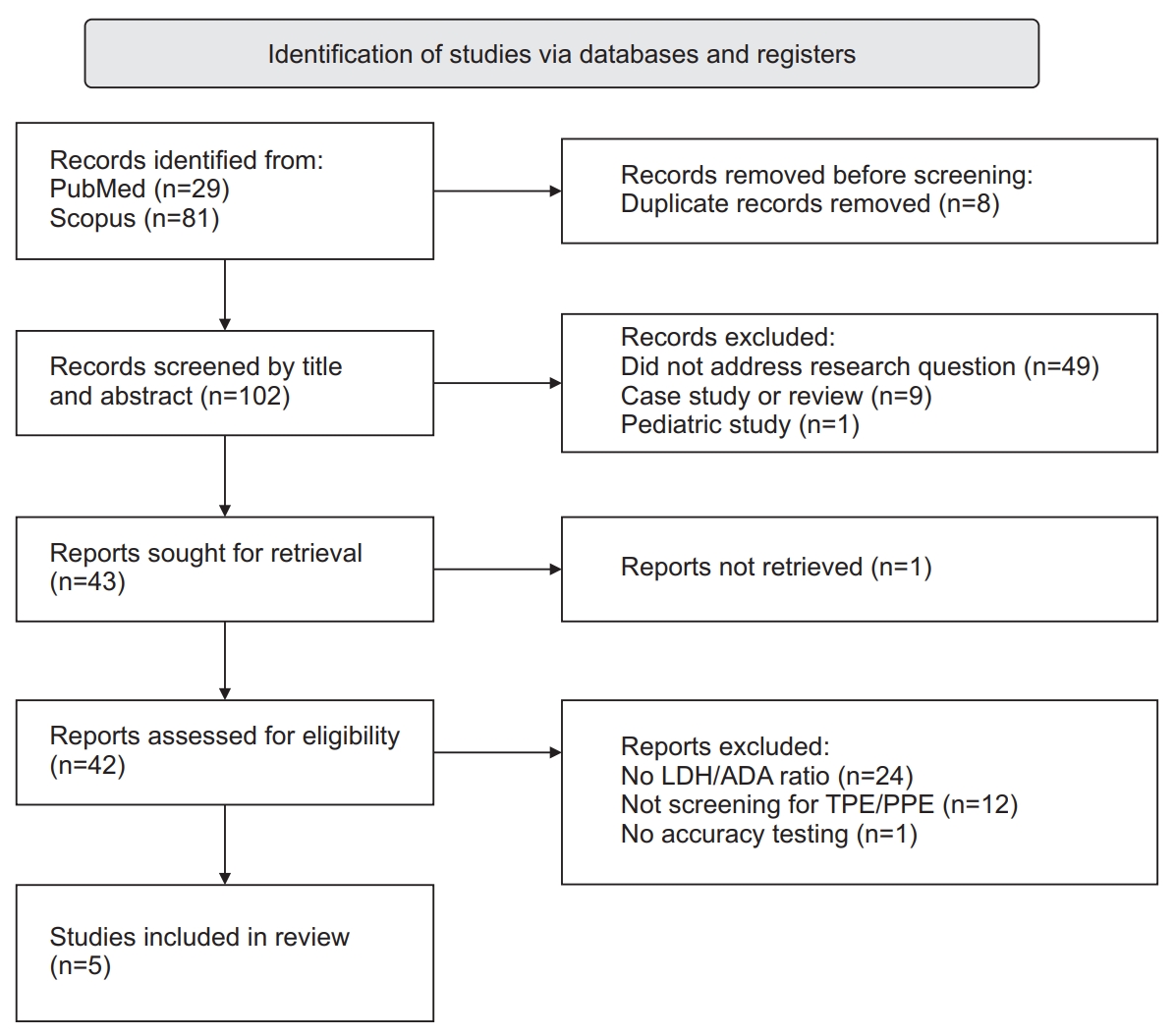
Based on the electronic database search, 110 studies were found, eight of which were excluded as duplicates (Figure 1). Fifty-nine studies were excluded based on their titles and abstracts. The remaining 43 studies were subjected to full-text assessment. One study was not retrievable. We excluded 35 of these studies because they did not mention the LDH/ADA ratio (n=24), of the absence of TPE or (PPE) (n=12), and the absence of accuracy testing (n=1). Finally, a total of five studies were deemed eligible for the systematic review, encompassing a total of 2,407 patients (Figure 1). These studies were published between 2017 and 2022. All of the five studies were observational, single-center studies, and only one was a prospective study [6]. The studies were conducted in South Africa, New Zealand, Taiwan, and China [4-6,11,12]. There were no randomized control trials. Of note, one of the studies only recruited patients with ADA value >40 U/L [12]. Criteria for TPE were consistent in all but one study, which only included TB culture-positive samples [11]. Meanwhile, criteria for PPE differed between studies; one study did not explicitly state the PPE criteria [6]. Variation in PPE criteria included a documented clinical diagnosis in the absence of alternative causes [11], positive pleural or sputum culture with preceding clinical pulmonary infection [12], and clinical pulmonary infection in the absence of TPE [4,5]. The characteristics of the included studies and their patients are presented in Table 1.
3. Role of LDH/ADA in differentiating PPE and TPE
Cited LDH/ADA ratio cutoff values for TPE differed in each study, ranging from <7.5 (sensitivity 64%, specificity 96%) in a South African cohort [6], to <25 (sensitivity 97%, specificity 62% in a South African cohort [6]; sensitivity 100%, specificity 61.6% in a New Zealand cohort [11]) PPV was much lower (ranging from 8.5 to 57.3 at cutoff values of <15 to <25, with ADA >15 or >30) but NPV was high (99.5 to 100) in the New Zealand cohort, where TB prevalence is very low [11]. Specific cutoff values for PPE were >14.5 (sensitivity 79.9%, specificity 78.5%) in the Taiwanese cohort [12]. LDH/ADA ratio was found to be superior to ADA alone in differentiating TPE from alternative diagnoses (area under the receiver operating characteristic curve [ROC], 0.92 [p<0.001] vs. 0.88 [p<0.001]) [6,12], while other studies demonstrated noninferiority [11]. However, the LDH/ADA ratio had less PPV and NPV than ADA alone in a setting of lymphocyte-predominant pleural effusion [6]. In studies that employed multiple cutoff values, PPV and sensitivity increased as the LDH/ADA ratio cutoff values decreased, while NPV and specificity decreased accordingly. LDH/ADA ratio is significantly lower in TPE than in PPE [5]. Detailed information can be found in Table 2.
4. Additional value of ADA or lymphocyte-predominance in combination with LDH/ADA ratio
When compared to LDH/ADA ratio alone, no difference was found with the combination of LDH/ADA ratio and lymphocyte-predominance, with the authors proposing that a ratio <7.5 precludes the need for fluid lymphocyte testing [6]. Combined LDH/ADA ratio and elevated fluid ADA increased specificity and PPV [11], while a combination of LDH/ADA, ADA, serum albumin, serum LDH, and pleural fluid LDH/total protein provided a 100% sensitivity and 98.7% specificity [4]. More details on this can be found in Table 2. Median values for ADA, LDH, and LDH/ADA ratios are presented in Table 3.
5. Risk of bias assessment (QUADAS-2)
Selection bias was high for all the studies identified, as convenience sampling was used in all five studies. None of the studies were randomized. A summary table of risk of bias assessments is described in detail in Supplementary Table S1.
6. Domain: patient selection
Patient selection was conducted via convenience sampling from a database or registry [4,6,12], lab samples [11], or consecutive patients presenting to a healthcare center [5]. In all studies, none of the sampling methods were randomized. Inclusion and exclusion criteria varied greatly between the studies. One study included all exudative pleural effusions [4], one study included both exudative and transudative effusions [11], one study included only effusions with ADA levels above 40 [12], and three studies were conducted on patients with an established diagnosis. Most studies excluded subjects with incomplete clinical data. Two studies did not define their exclusion criteria [4,5]. One study excluded undiagnosed exudates [6]. The risk of bias for patient selection was high in all studies and may have led to underestimation of diagnostic accuracy.
7. Domain: index test
In all studies, the LDH/ADA ratio was interpreted with knowledge of the reference standards, as they assessed patients with a known diagnosis based on reference standard to compare their respective LDH/ADA ratios [4-6,11,12]. The LDH/ADA ratio was assessed retrospectively after the diagnosis was made; hence, there was a high risk of bias. Of note, the studies had varying analysis groups. One study subclassified PPE into uncomplicated and complicated groups and included two categories that were not found in other studiesŌĆö connective tissue disease-related effusion and chronic, nonspecific pleuritis [4]. A few studies had preset LDH/ADA ratio thresholds. The New Zealand cohort used a ratio <25 and <15; these values were determined based on historical data from an internal audit conducted at the study site [11], but the threshold was determined after data collection. ROC curves were used to identify the optimal cutoff points in other studies [4-6,12]. Two studies excluded patients with an ADA level of below 40 [12]. This may have led to overestimated test performance.
8. Reference standard
PPE was explicitly defined in all but one study [6]. Meanwhile, pleural TB was consistently defined in all studies. The authors have low concerns regarding both whether the reference standard is likely to correctly classify the target condition and whether the target condition will not match the review question. This is because, in all studies, LDH/ADA ratios were calculated and interpreted retrospectively in patients with the already known diagnoses; hence, there is no risk of bias.
9. Flow and timing
There were no concerns regarding verification bias, as all studies retrieved ADA and LDH samples in the same pleural fluid sampling time frame. In one study only 68.7% of the pleural fluid samples were cultured for TB, as the practice in the center was only to culture those with ADA >15 [11]. A few studies had significant exclusion of patients. In one study, 39 out of 267 patients were excluded from analysis due to missing data (n=24) or undiagnosed effusion (n=15) [6]. In another study, 42 out of 353 patients were excluded from analysis due to missing data [12]. Some studies had no described exclusions while two studies mentioned excluding patients who did not fulfill inclusion criteria but did not mention how many patients were excluded [4]. A summary of all studies is presented in Table 1.
Discussion
Overall, the studies demonstrated high heterogeneity in terms of methodology and clinical characteristics. Heterogeneity was statistically significant as well, as evidenced by a chi-square value for between studies heterogeneity of 140.59 (p<0.001) for sensitivity and 1,226.87 (p<0.001) for specificity (data not shown). Furthermore, using I2 statistics, heterogeneity was also quantified to be 87.9% and 98.6%, respectively (data not shown). We considered that even if statistical heterogeneity were to be improved by subgroup analyses, the vastly different and heterogeneous nature of the methodologies and clinical characteristics of the studies rendered meta-analysis fundamentally unsuitable and invalid. Thus, meta-analysis was not done and is not presented in this study.
While data analysis suggests that the pleural fluid LDH/ADA ratio is significantly lower in TPE compared to PPE, the optimal cutoff value to differentiate TPE and PPE remains undefined and is most likely influenced by various confounding factors. Various ratios of between 14.2 to 16.2 have been suggested, each with varying levels of sensitivity and specificity. Potential causes of heterogeneity in cutoff values include differing study designs and laboratory thresholds or clinical factors, which may be summed up by the agent-host-environment triad, whereby agent factors include TB incidence and strain, host factors include genetic variability and susceptibility, and environment factors include climate, living conditions, and geographic differences. TB incidences differ the most between Beukes et al. [6] and Blakiston et al. [11] (TB incidence of >500/100,000 in South Africa vs. 6.4/100,000 in New Zealand), while for the three other studies, TB incidences ranged from 21.7 to 103.5/100,000 [4,5,12-15]. In areas with low TB burden, pleural fluid LDH/ADA ratio cutoff value with a high NPV might be sufficient to rule-out TPE. However, this broad interpretation does not account for the variable thresholds used in different individual studies. To further complicate matters, it is known that different ADA assays may cause slightly different resultsŌĆöfor example, the Giusti method has a positive bias compared to the non-Giusti Diazyme method [16].
Although the current strength of evidence is weak, a low pleural fluid LDH/ADA ratio may alert the astute clinician to the diagnosis of TPE. Therefore, it is probably appropriate to consider empirical treatment for TPE in these patients, especially in high TB prevalence areas, and if other clinical, radiologic, and laboratory parameters are consistent with TPE. This option is attractive in resource-limited settings, among patients who were not keen on further invasive tests, or among patients with small amounts of pleural effusion or dense septations rendering medical thoracoscopy difficult to perform. Nevertheless, it is vital to exercise caution due to the chances of misclassifying non-TPE patients among patients with true TPE. The availability of pleural fluid ADA should not deter healthcare professionals from offering other standard-of-care tests such as AbramŌĆÖs needle pleural biopsy or medical thoracoscopy whenever available. The current evidence is insufficient to recommend the use of the LDH/ADA ratio for neutrophil-predominant exudative effusion, and further studies are needed in this area. Other parameters like the pleural fluid ADA/serum C-reactive protein ratio may facilitate differentiating between TPE and PPE [17].
Data from the two largest studies, which also encompass two geographic areas of highest and lowest TB prevalence (South Africa and New Zealand, respectively), out of the five studies seem to suggest an LDH/ADA ratio cutoff of <15 as having the optimal PPV to differentiate TPE from PPE [6,11]. One caveat to this recommendation is that PPV is optimal at an ADA value of >30 [11]. However, this recommendation is not meant to be intransigent; moving forward, studies with more rigorous methodology and larger sample sizes should be conducted. It would not be surprising to have geographic-specific or population-specific values in the future.
LDH/ADA ratio value is probably <15 (we all need to agree on this). In the Beukes study (highest TB incidence) PPV is 92.0 and NPV is 79.0, and in the Blakiston study (lowest TB incidence) PPV is 17.3 and NPV is 99.5 (PPV improved significantly if LDH/ADA paired with ADA >30). Moving forward, more studies with better methodological quality and larger study populations should be conducted.
Our systematic review is not without limitations. First, the analysis was restricted to articles published in English language, thus non-English studies may have been missed out. The quality of most studies was suboptimal owing to their retrospective, single-center design. None of the studies were randomized. Also, the differing TB incidences suggest differing prevalence and by extension pretest probabilities, affecting PPVs and NPVs. Although sensitivity and specificity are the inherent values of the tool itself and not affected by disease prevalence, the utility of the LDH/ADA ratio to differentiate between TPE and PPE should be viewed from the perspective of clinical decision-making, and thus robust PPV and NPV values are more useful. This accentuates the limitation further. Several biases may have occurred in the studies included in our review, such as confirmation bias due to diagnosis or suspicion of TB being influenced by ADA results. One study included patients with transudative effusions, which despite potentially improving specificity estimates, would contribute to increased heterogeneity [11]. Last but not least, none of the studies included in our review performed external or temporal validation.
In conclusion, LDH/ADA ratio is significantly lower in TPE compared to PPE, and may be employed in clinical encounters where medical thoracoscopy is contraindicated or unavailable, or when emergent decisions on commencement of antituberculous treatment are warranted due to clinical urgency or public health concerns, such as outbreaks. Contemporary evidence suggests that an LDH/ADA ratio cutoff value of <15 appears useful in differentiating TPE and PPE. Nevertheless, the interpretation of the LDH/ADA ratio requires the clinician to evaluate the pretest probability of TB. Rigorous studies with improved designs integrating randomization, standardization of ADA assays, and studies that limit biases, are needed in the future.
 PDF Links
 PubReader
 ePub Link
 Data Sharing Statement
 Full text via DOI
 Supplement
 Print
Print Download Citation


