Tuberc Respir Dis > Volume 87(3); 2024 > Article
Kim, Moon, Min, and Lee: Proposed Etiotypes for Chronic Obstructive Pulmonary Disease: Controversial Issues

Abstract

The 2023 Global Initiative for Chronic Obstructive Lung Disease (GOLD) revised the definition of chronic obstructive pulmonary disease (COPD) to broadly include a variety of etiologies. A new taxonomy, composed of etiotypes, aims to highlight the heterogeneity in causes and pathogenesis of COPD, allowing more personalized management strategies and emphasizing the need for targeted research to understand and manage COPD better. However, controversy arises with including some diseases under the umbrella term of COPD, as their clinical presentations and treatments differ from classical COPD, which is smoking-related. COPD due to infection (COPD-I) and COPD due to environmental exposure (COPD-P) are classifications within the new taxonomy. Some disease entities in these categories show distinct clinical features and may not benefit from conventional COPD treatments, raising questions about their classification as COPD subtypes. There is also controversy regarding whether bronchiectasis with airflow limitations should be classified as an etiotype of COPD. This article discusses controversial issues associated with the proposed etiotypes for COPD in terms of COPD-I, COPD-P, and bronchiectasis. While the updated COPD definition by GOLD 2023 is a major step towards recognizing the disease’s complexity, it also raises questions about the classification of related respiratory conditions. This highlights the need for further research to improve our understanding and approach to COPD management.

Key Figure

Introduction

The Global Initiative for Chronic Obstructive Lung Disease (GOLD) 2023 revised the definition of chronic obstructive pulmonary disease (COPD) and suggested a newly defined taxonomy, i.e., etiotypes [1]. Before GOLD 2023, the COPD definition had the following three components: (1) respiratory symptoms and airflow limitation; (2) airway and/or alveolar abnormalities usually caused by significant exposure to noxious particles or gases; and (3) host factors, including abnormal lung development [2]. In 2023, GOLD emphasized “heterogenous lung conditions” to embrace various causes of COPD. The main goal of this change was to uncover the heterogeneity of etiologies and pathogenesis of COPD [3]. This will allow us to provide tailored strategies to reduce exposure to various risk factors for COPD and facilitate research to reveal the heterogenous natural course of COPD according to each etiotype.
Despite the considerable disease burden, only a small percentage of individuals with COPD are diagnosed and receive appropriate care [4]. COPD disproportionately affects people in low- and middle-income countries, mostly located in Asia and Africa [5]. While tobacco smoking is the leading risk factor for COPD [6], there are many other contributing factors, such as air pollution, occupational exposure to dust and fumes, and respiratory infections [7,8]. By expanding the definition of COPD to include a broader range of heterogeneous lung conditions, it is expected that clinicians will be able to identify more individuals with COPD earlier and provide them with the appropriate preventive measures and/or treatment before irreversible damage occurs. However, there is concern about whether COPD could be used as an ambiguous umbrella term encompassing various disease entities [9]. In this review, we discuss newly proposed etiotypes of COPD and related controversy.

Change in COPD Definition and Newly Defined Taxonomy

1. History of COPD definition

Changes in major components of COPD from 2011 to 2023 are presented in Table 1. Historically, COPD has been defined as a condition characterized by persistent airflow limitation linked to noxious particles or gases [2]. However, the 2023 GOLD report updated the COPD definition to expand the boundaries of the disease in terms of etiologies [1].
Although the proportion of non-smoking-related COPD is higher than smoking-related COPD globally [5,10], COPD has been generally regarded as a smoking-related disease [8] since the development of major COPD research and clinical guidelines has mainly been driven by Western academic societies. Accordingly, COPD caused by infections and air pollution, which are more common in low-to-middle income countries, including many Asian countries, has been largely neglected by the research community. Accordingly, the previous definitions before GOLD 2023 did not fully account for these individuals. From this perspective, the revised definition of COPD seems to better define COPD by reflecting broader situations across the world.

2. Proposed taxonomy for COPD

The updated definition of COPD has led to the need for a new classification system. The GOLD committee has proposed a taxonomy for COPD that focuses on the underlying causes of the disease (Table 2). However, this definition may cause controversy since there have been no in-depth considerations for pathogenesis and clinical presentations. Some disease entities traditionally regarded as diseases distinguishable from COPD may now be included as a subtype of COPD.

Controversial Issues 1: Bronchiectasis with Airflow Limitation

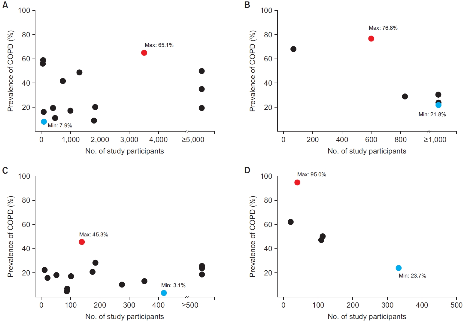
Bronchiectasis is a disease entity of clinical and radiological diagnosis (abnormal dilatation of the bronchi), characterized by cough, sputum production, and recurrent respiratory infections that overlap with other airway diseases [11-13]. The overlap between bronchiectasis and COPD is very common. A large proportion of individuals with COPD have bronchiectasis on chest computed tomography (CT) [14]. Also, a high proportion of individuals with bronchiectasis have airflow limitation or COPD (Figure 1) [15,16].
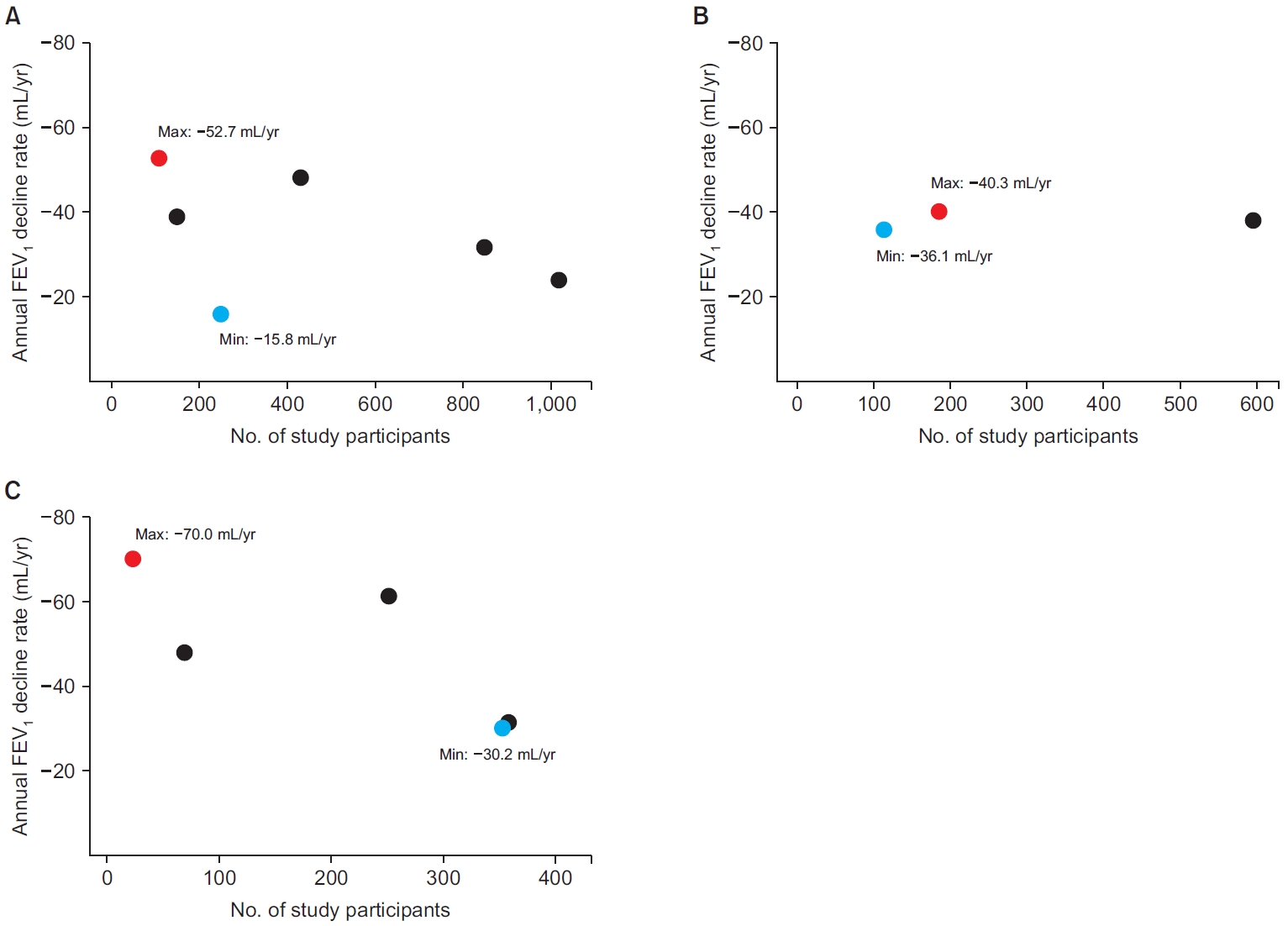
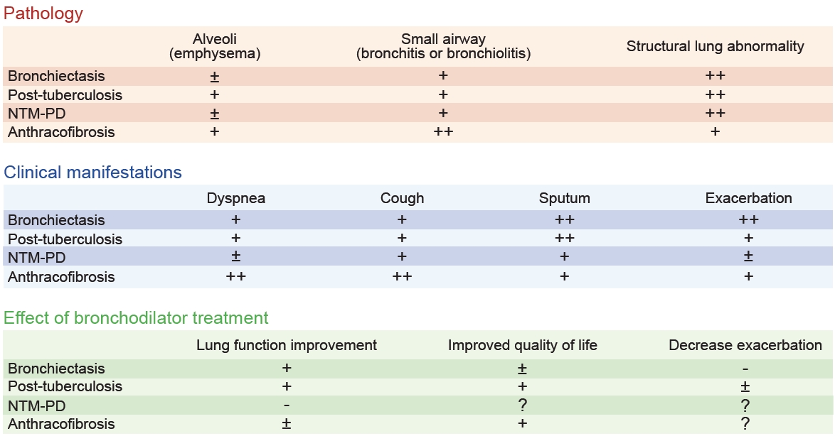
Despite a considerable overlap between the two diseases, bronchiectasis has been traditionally considered a distinct disease entity from COPD. Supporting this position, the GOLD committee mentioned that calling bronchiectasis an “etiotype” of COPD is incorrect since bronchiectasis has many causative etiologies and its own nosological entity [17], concluding that bronchiectasis represents an important comorbidity of COPD that should be identified, not an etiotype of COPD. The pathology of bronchiectasis is also different from classical COPD in terms of major involvement sites. While classical COPD is characterized by alveoli involvement and/or small airway involvement [18], the structural dilatation of bronchi is the main characteristic of bronchiectasis, although bronchiectasis is often accompanied by bronchiolitis or bronchiolitis (Table 3) [19]. Rapid lung function decline is an important pathophysiology leading to COPD. However, changes in lung function in bronchiectasis vary widely across studies (Figure 2) [20-25].
Furthermore, the main treatment strategies of bronchiectasis are also different from those of COPD, although the bronchiectasis treatment guidelines recommend using a bronchodilator in individuals with coexistence of bronchiectasis and COPD [26]. While airway clearance techniques are the cornerstone therapy of bronchiectasis treatment, the use of bronchodilators is the central management strategy for COPD [26]. Some studies have suggested that bronchodilators can be effective in improving lung function in bronchiectasis [27-29]. Although a randomized controlled trial for dual bronchodilators in bronchiectasis is ongoing [30], the effect of bronchodilators on the treatment outcomes of bronchiectasis has not been established other than lung function (e.g., quality of life and acute exacerbations) (Table 3) [31,32]. For example, although inhaled tiotropium increased lung function in individuals with bronchiectasis and airflow obstruction, it did not show reduced exacerbation or improved quality of life [27], which are all observed in classical COPD [33]. Exacerbation management is also different. In bronchiectasis, antibiotics are the main treatment, and systemic steroids are not routinely recommended [26], while in COPD, a combination of bronchodilator, systemic steroid, or antibiotics is used [8].
Despite these recommendations, a considerable proportion of experts on airway diseases suggest that bronchiectasis is an etiotype of COPD. For example, a survey of Korean experts showed that 53% of survey participants agreed with the following statement: “The definition of COPD includes patients with persistent airflow obstruction due to bronchiectasis, even without previous exposure to gases and dust, including cigarette smoking” (note: bronchiectasis mentioned here refers to patients with a main diagnosis of bronchiectasis for their respiratory condition and treatment) [34]. Although the reasons for their agreement on the statement were not provided, it seems that the experts providing agreement on the statement would have focused on the development of persistent airflow limitation and improved lung function in bronchiectasis following bronchodilator treatments [27-29].
Most studies are needed to clarify the natural course of the overlap of bronchiectasis and COPD. The coexistence of bronchiectasis and COPD leads to a higher disease burden [35], but there is currently no standardized definition for this population. Fortunately, a Delphi consensus definition for bronchiectasis-COPD overlap has recently been suggested by the European Multicentre Bronchiectasis Audit and Research Collaboration (EMBARC) Airways Working Group, which includes Radiological, Obstruction, Symptoms, and Exposure (ROSE) criteria [36]. The ROSE criteria consist of: (1) Radiological (abnormal bronchial dilatation in one or more pulmonary segments in more than one lobe and radiological findings of airway visible within 1 cm of pleura and/or lack of tapering sign); (2) Obstruction (a functional obstructive pattern [post-bronchodilator forced expiratory volume in 1 second (FEV1)/forced vital capacity <0.7]); (3) Symptoms (two or more of the following symptoms [cough, expectoration, dyspnea, fatigue, and frequent lower airway infections]); (4) Exposure (ever smoker with more than 10 pack-years or other toxic exposure). Future studies using this definition would provide more evidence of the relationship between the two diseases while reducing the heterogeneity of clinical characteristics of the study population caused by different definitions of bronchiectasis-COPD overlap.

Controversial Issues 2: COPD Due to Infection

In the revised GOLD 2023 guideline, childhood infection, tuberculosis (TB), and human immunodeficiency virus (HIV)-associated airflow limitation are proposed as examples of COPD due to infection (COPD-I). Traditionally, obstructive lung disease caused by those diseases was not classified as COPD, even though infection-related obstructive lung diseases are frequently encountered in real-world clinics. With accumulating data on the association between these three infectious conditions and COPD, classifying this disease entity into COPD-I is reasonable, and it is welcome news that we now have a rationale on which to base various preventive modalities and treatments for individuals with COPD-I. For tuberculosis-associated COPD, we focus on the accumulated evidence on the relationship between COPD and TB rather than a controversial issue. We will discuss controversial issues on COPD related to other respiratory infectious conditions (e.g., non-tuberculous mycobacterial pulmonary disease [NTM-PD]).

1. Tuberculosis-associated COPD

Pulmonary TB is an important cause of airflow limitation in Korea; Rhee et al. [37] found that approximately 75% of individuals with a TB-destroyed lung (TBDL) had airflow limitation. Another Korean study also showed that evidence of previous TB on chest X-ray was associated with increased odds of airflow limitation even after adjustment or exclusion of smokers [38]. Regarding the overall risk of COPD in post-TB individuals, a meta-analysis of studies evaluating the association between pulmonary TB and the risk of COPD found that individuals with a history of pulmonary TB had a 2.59-fold increased risk of developing COPD [39]. In studies using the Korean National Health Examination Survey data, a history of pulmonary TB was frequently observed in individuals with airflow limitation [40,41]. Likewise, COPD is prevalent in the post-TB cohort [38,42,43]. While the size and enrollment criteria for subjects who have experienced pulmonary TB vary across studies, the prevalence of COPD in individuals with a history of pulmonary TB ranges from 21.8% to 76.8%, indicating a close connection between both conditions (Figure 1) [37,44-48]. Also, a relatively consistent and fast decline in lung function of about -40 mL/year in FEV1 has been reported post-TB individuals [37,49,50], which is higher than the FEV1 decline in Korean healthy never smokers (-31.8 mL/year) [51].
Previous COPD definitions, including a history of exposure to noxious particles as a criterion, led to a debate about whether airflow limitation observed in post-TB individuals can be regarded as COPD. However, growing evidence shows that post-TB with airflow limitation could be classified as COPD-I. Park et al. [52] investigated the impact of a history of pulmonary TB on the severity and outcomes of COPD using the Korean COPD cohort. The prior TB group had worse respiratory symptoms and quality of life, lower lung function, and increased prevalence of exacerbation compared to the non-TB group. Pathology also supports that post-TB airflow limitation should be classified as COPD-I because they have alveoli involvement as well as small airway pathologies (Table 3) [53]. Additionally, bronchodilators, a cornerstone therapy for COPD, significantly prevented future lung function decline and improved COPD outcomes in individuals with COPD accompanied by TBDL [54]. Although the prevention effect of bronchodilators for exacerbation was unclear, using bronchodilators was also associated with a decrease in mortality among this population (Table 3) [55,56]. These findings align with the new concept that post-TB-related airflow limitation may be a distinct subtype of COPD.

2. Other respiratory infections: NTM-PD

Certain chronic respiratory infections other than pulmonary TB may be linked with COPD, indicating the potential existence of COPD-I other than COPD related to childhood infection, HIV infection, and TB. Although other infection-related airflow limitation were not included in the guideline, the GOLD committee commented that various infections could be causes for COPD development, and NTM-PD is one such candidate [17]. NTM-PD is a chronic infectious lung disease characterized by its persistence and slow-progressing nature, with its prevalence and disease burdens steadily rising [57]. The airflow limitation is frequently encountered in the management of NTM-PD. In a study of 358 individuals with NTM-PD, 8.2% of subjects experienced a change in lung function from normal to obstructive during a median 5.6-year follow-up [58]. In the NTM studies evaluating COPD as a comorbidity, the comorbid COPD was reported up to 65.7% in individuals with NTM-PD [59-73], suggesting a comparable prevalence of COPD within individuals with TBDL (Figure 1). In contrast, NTM-PD was also found in individuals with COPD, and comorbid NTM-PD is associated with poor prognosis [74,75]. Moreover, the decline in lung function is often pronounced in individuals with NTM-PD (Figure 2) [58,75-78]. As in TB, NTM-PD shares common features of pathologic changes in COPD, such as alveoli involvement and small airway involvement [79]. However, information on the treatment response to bronchodilators is scarce in individuals with NTM-PD (Table 3).
There is very little literature that highlights the underlying mechanism linking COPD and NTM-PD. There may be a bidirectional association between these two diseases, promoting the development of each condition by the other condition. NTM-associated airway inflammation may cause small airway obstructions [80], and in individuals with COPD, defense against the pathogen is reduced [81]. Nevertheless, there are distinguishing features in NTM-PD associated with airflow limitation from classical COPD. First, antimicrobial agents remain the main treatment for NTM-PD [82]. The role of bronchodilators has not been established in NTM-PD, even when the individuals have airflow limitation [58]. Additionally, airflow limitation might be improved through appropriate infection control [83]. Second, considering these chronic respiratory infections commonly exist as a comorbid condition of structural lung disease, most airflow limitation in these populations may be associated with underlying lung parenchymal destruction [28]. Third, the natural course and clinical outcomes are mainly driven by the features of NTM (NTM species, cavity, acid-fast bacilli smear positivity, etc.) rather than those of COPD [84].
In this context, COPD may be an under-recognized condition in the management of NTM-PD, but it is uncertain whether NTM-PD could be classified as COPD-I. More studies will be required to classify NTM-PD-associated airflow limitation as COPD-I.

Controversial Issues 3: COPD Due to Environmental Exposure

1. Bronchial anthracofibrosis

Bronchial anthracofibrosis (BAF) is a chronic lung disease caused by the inhalation of fine particles of carbon from biomass fuel smoke [85]. BAF is commonly found in elderly women who are never smokers and live in low- and middle-income countries where biomass fuels are commonly used for cooking and heating [86]. Also, several studies reported the association between pulmonary TB and BAF [85]. This disease entity is often diagnosed as asthma or COPD because of similar spirometric findings, i.e., obstructive spirometry pattern [87].
The prevalence of airflow limitation in BAF varies widely; most studies reporting spirometric results in BAF were conducted based on a relatively small number of individuals, and the prevalence of airflow limitation was 24% to 95% (Figure 1) [88-90]. In Korea, the rate of having BAF in COPD seems relatively frequent. According to a single-center respective study in Korea, one-fourth of the individuals had a BAF based on CT criteria among the those with COPD exacerbation [87]. From a pathologic view, BAF and COPD both commonly affect small airways despite their distinct patterns of pathological changes (Table 3) [91].
However, some features of BAF are different from those of classical COPD. First, although not frequent, BAF occasionally involves larger airways and regional mediastinal lymph nodes, which leads to mechanically obstructive lung disease rather than a small airway disease. In those cases, bronchoscopic intervention (e.g., mechanical dilatation of a large airway) has a central role in the treatment of BAF [92]. Second, the effects of bronchodilators are less significant than those in individuals with COPD. While some research demonstrated the effect of bronchodilators, unresponsiveness to bronchodilators is more commonly reported (Table 3) [93,94]. Therefore, more studies are needed on whether this condition can be classified as COPD-P.

2. Pneumoconiosis

COPD frequently coexists with pneumoconiosis, raising the question of the prevalence of COPD in individuals who suffer from pneumoconiosis. Several studies reported a common occurrence of airflow limitation, suggesting COPD in individuals with pneumoconiosis, with the prevalence of COPD being around 20% [95]. In light of the latest taxonomy for COPD, such cases might be classified as COPD-P. However, some issues should be considered before classifying pneumoconiosis as a COPD-P. First, pneumoconiosis has a distinctive clinical manifestation, with progressive massive fibrosis being the most prominent example [95]. Progressive massive fibrosis is a severe form of pneumoconiosis where large amounts of the lung tissue turn fibrous, impairing its function [96], which is different from classical COPD. Second, the underlying pathology of lung dysfunction in pneumoconiosis may differ from that of classical COPD. Pneumoconiosis is often a result of prolonged exposure to relatively large dust particles, leading to specific structural changes in the lungs, which is distinguished from the small airway dysfunction in COPD [97]. Lastly, pneumoconiosis is not a single disease entity. There is heterogeneity among diseases in the pneumoconiosis category [97]. Besides, the impact of bronchodilator treatment on the treatment outcomes in individuals with pneumoconiosis-related airflow limitations has not been studied.

Conclusion

The 2023 GOLD Committee revised the definition of COPD to reflect its heterogeneity of etiologies and pathogenesis and to enable tailored strategies reducing exposure to COPD risk factors across various etiotypes. Accumulating evidence agrees with the suggestion by the GOLD committee that TB is an important etiology of COPD-I. However, there may be controversy in classifying some diseases, such as bronchiectasis, respiratory infections other than TB, BAF, and pneumoconiosis, as etiotypes of COPD. Future research is warranted to clarify the role of these diseases in COPD development and progression.

Notes

Authors’ Contributions

Conceptualization: Min KH, Lee H. Methodology: Lee H. Investigation: Kim SH. Writing - original draft preparation: Kim SH, Moon JY. Writing - review and editing: all authors. Approval of final manuscript: all authors.

Conflicts of Interest

Sang Hyuk Kim is an early career editorial board member and Ji-Yong Moon and Kyung Hoon Min are editors of the journal, but they were not involved in the peer reviewer selection, evaluation, or decision process of this article. No other potential conflicts of interest.

Funding

No funding to declare.

Fig. 1.
Prevalence of chronic obstructive pulmonary disease (COPD). (A) Bronchiectasis. (B) Tuberculosis-destroyed lung. (C) Non-tuberculous mycobacterial pulmonary disease. (D) Anthracofibrosis.
trd-2023-0194f1.jpg
Fig. 2.
Annual decline in lung function. (A) Bronchiectasis. (B) Tuberculosis-destroyed lung. (C) Non-tuberculous mycobacterial pulmonary disease. There have been no research results on changes in lung function in anthracofibrosis. FEV1: forced expiratory volume in 1 second.
trd-2023-0194f2.jpg
trd-2023-0194f3.jpg
Table 1.
Changes to major components of COPD definition
Year 2011-2016 2017-2019 2020-2022 2023
Definition COPD, a common preventable and treatable disease, is characterized by persistent airflow limitation that is usually progressive and associated with an enhanced chronic inflammatory response in airways and the lungs to noxious particles or gases. Exacerbations and comorbidities contribute to the overall severity in individual patients. COPD is a common, preventable and treatable disease that is characterized by persistent respiratory symptoms and airflow limitation that is due to airway and/or alveolar abnormalities usually caused by significant exposure to noxious particles or gases. COPD is a common, preventable, and treatable disease that is characterized by persistent respiratory symptoms and airflow limitation that is due to airway and/or alveolar abnormalities usually caused by significant exposure to noxious particles or gases and influenced by host factors, including abnormal lung development. Significant comorbidities may have an impact on morbidity and mortality. COPD is a heterogeneous lung condition characterized by chronic respiratory symptoms (dyspnea, cough, sputum production and/or exacerbations) due to abnormalities of the airway (bronchitis, bronchiolitis) and/or alveoli (emphysema) that cause persistent, often progressive, airflow obstruction.
Symptoms Persistent respiratory symptoms Persistent respiratory symptoms Chronic respiratory symptoms (dyspnea, cough, sputum and/or exacerbations)
Exposure Noxious particles or gases Noxious particles or gases Noxious particles or gases Not included in the definition, but various etiotypes were separately mentioned.
Pathology Chronic inflammatory response in airway and the lungs Airway and/or alveolar abnormalities Airway and/or alveolar abnormalities Abnormalities of the airway (bronchitis, bronchiolitis) and/or alveoli (emphysema)
Physiology Persistent AFL Persistent AFL Persistent AFL Persistent, often progressive airway obstruction

COPD: chronic obstructive pulmonary disease; AFL: airflow limitation.

Table 2.
Proposed classification and taxonomy for COPD
Etiotypes Classification Details Description
Type 1: genetically determined COPD COPD-G Alpha-1-antitrypsin deficiency Alpha-1-antitrypsin deficiency and other genetic variants with smaller effects acting in combination
Telomerase reverse transcriptase mutations
Other genetic variants
Type 2: COPD related to early life events COPD due to abnormal lung development (COPD-D), COPD, and asthma (COPD-A) Prematurity (chronic lung disease of prematurity, bronchopulmonary dysplasia) Early life events (including premature birth and low birth weight, among others) and childhood asthma
Childhood asthma
Type 3: infection-related COPD COPD-I Childhood respiratory infections COPD that is associated with early life or particular infections (pulmonary tuberculosis and HIV)
Tuberculosis-associated COPD
HIV-associated COPD
Type 4: COPD related to smoking or vaping COPD-C Tobacco smoking Exposure to tobacco smoke (including in utero), passive smoking, vaping, e-cigarette, or cannabis
In utero exposure to tobacco smoke
Passive smoking
Vaping or e-cigarette smoking
Cannabis smoking
Type 5: environmental exposure-related COPD COPD-P Exposure to indoor air pollutants Exposure to household pollution, ambient air pollution, wildfire smoke, and occupational hazards
Outdoor air pollution and smog
Wildfire smoke
Occupational exposures
Others COPD of unknown cause (COPD-U)

COPD: chronic obstructive pulmonary disease; HIV: human immunodeficiency virus.

Table 3.
Pathology, chronic manifestations, and effect of bronchodilator treatment
Variable Bronchiectasis Post-tuberculosis NTM-PD Anthracofibrosis
Pathology
 Alveoli (emphysema) ± + ± +
 Small airway (bronchitis or bronchiolitis) + + + ++
 Structural lung abnormality ++ ++ ++ +
 Reference [11,17,18] [47,48] [58,69,73] [85,86]
Clinical manifestations
 Dyspnea + + ± ++
 Cough + + + ++
 Sputum ++ ++ + +
 Exacerbation ++ + ± +
 Reference [11,19] [28,37,46] [58,69] [80,82,84,88]
Effect of bronchodilator treatment
 Lung function improvement + + - ±
 Improved quality of life ± + ? +
 Decrease exacerbation - ± ? ?
 Reference [19-22] [48-50] [52] [87,88]

The symbols were determined based on: (?) no study exists, (-) only negative studies exist, (±) conflicting result studies exist, (+) some positive studies exist, (++) several positive studies exist.

NTM-PD: non-tuberculous mycobacterial pulmonary disease.

REFERENCES

1. Global Initiative for Chronic Obstructive Lung Disease (GOLD). Global strategy for prevention, diagnosis and management of COPD: 2023 report [Internet]. Fontana: GOLD; 2023 [cited 2024 Feb 16]. Available from: http://goldcopd.org/2023-gold-report-2.

2. Choi JY, Rhee CK. Diagnosis and treatment of early chronic obstructive lung disease (COPD). J Clin Med 2020;9:3426.
crossref pmid pmc
3. Stolz D, Mkorombindo T, Schumann DM, Agusti A, Ash SY, Bafadhel M, et al. Towards the elimination of chronic obstructive pulmonary disease: a Lancet Commission. Lancet 2022;400:921-72.
crossref pmid pmc
4. Yoo KH, Kim YS, Sheen SS, Park JH, Hwang YI, Kim SH, et al. Prevalence of chronic obstructive pulmonary disease in Korea: the fourth Korean National Health and Nutrition Examination Survey, 2008. Respirology 2011;16:659-65.
crossref pmid
5. Adeloye D, Song P, Zhu Y, Campbell H, Sheikh A, Rudan I, et al. Global, regional, and national prevalence of, and risk factors for, chronic obstructive pulmonary disease (COPD) in 2019: a systematic review and modelling analysis. Lancet Respir Med 2022;10:447-58.
crossref pmid pmc
6. Bhatt SP, Kim YI, Harrington KF, Hokanson JE, Lutz SM, Cho MH, et al. Smoking duration alone provides stronger risk estimates of chronic obstructive pulmonary disease than pack-years. Thorax 2018;73:414-21.
crossref pmid pmc
7. Sarkar C, Zhang B, Ni M, Kumari S, Bauermeister S, Gallacher J, et al. Environmental correlates of chronic obstructive pulmonary disease in 96 779 participants from the UK Biobank: a cross-sectional, observational study. Lancet Planet Health 2019;3:e478-90.
crossref pmid
8. Christenson SA, Smith BM, Bafadhel M, Putcha N. Chronic obstructive pulmonary disease. Lancet 2022;399:2227-42.
crossref pmid
9. Lee H, Shin SH, Park HY, Lim SY. Can we call all obstructive lung diseases COPD? Eur Respir J 2023;61:2300462.
crossref pmid
10. Terzikhan N, Verhamme KM, Hofman A, Stricker BH, Brusselle GG, Lahousse L. Prevalence and incidence of COPD in smokers and non-smokers: the Rotterdam Study. Eur J Epidemiol 2016;31:785-92.
crossref pmid pmc pdf
11. O’Donnell AE. Bronchiectasis: a clinical review. N Engl J Med 2022;387:533-45.
crossref pmid
12. Moon SM, Choi H, Kang HK, Lee SW, Sim YS, Park HY, et al. Impacts of asthma in patients with bronchiectasis: findings from the KMBARC Registry. Allergy Asthma Immunol Res 2023;15:83-93.
crossref pmid pmc pdf
13. Kim SH, Kim C, Jeong I, Lee SJ, Kim TH, Lee CY, et al. Chronic obstructive pulmonary disease is associated with decreased quality of life in bronchiectasis patients: findings from the KMBARC Registry. Front Med (Lausanne) 2021;8:722124.
crossref pmid pmc
14. Hurst JR, Elborn JS, De Soyza A; BRONCH-UK Consortium. COPD-bronchiectasis overlap syndrome. Eur Respir J 2015;45:310-3.
crossref pmid
15. Marsland I, Sobala R, De Soyza A, Witham M. Multimorbidity in bronchiectasis: a systematic scoping review. ERJ Open Res 2023;9:00296-2022.
crossref pmid pmc
16. Kim SH, Lee H, Kim Y. Health-related quality of life after pulmonary tuberculosis in South Korea: analysis from the Korea National Health and Nutrition Examination Survey between 2010 and 2018. Health Qual Life Outcomes 2021;19:195.
crossref pmid pmc pdf
17. Agusti A, Anzueto A, Celli BR, Mortimer K, Salvi S, Vogelmeier CF, et al. GOLD 2023 executive summary: responses from the GOLD Scientific Committee. Eur Respir J 2023;61:2300616.
crossref pmid pmc
18. Higham A, Quinn AM, Cancado JE, Singh D. The pathology of small airways disease in COPD: historical aspects and future directions. Respir Res 2019;20:49.
crossref pmid pmc pdf
19. Akatli AN, Ulutas H, Turkmen Samdanci E, Celik MR. Bronchiectasis: retrospective analysis of clinical and pathological findings in a tertiary-care hospital. Int J Clin Pract 2022;2022:8773204.
crossref pmid pmc pdf
20. Borekci S, Gundogdu S, Harbiyeli DO, Musellim B. Annual FEV1 loss in patients with noncystic fibrosis bronchiectasis and affecting factors. South Med J 2022;115:328-32.
crossref pmid
21. Chalmers JD, Moffitt KL, Suarez-Cuartin G, Sibila O, Finch S, Furrie E, et al. Neutrophil elastase activity is associated with exacerbations and lung function decline in bronchiectasis. Am J Respir Crit Care Med 2017;195:1384-93.
crossref pmid pmc
22. Dhar R, Singh S, Talwar D, Murali Mohan BV, Tripathi SK, Swarnakar R, et al. Clinical outcomes of bronchiectasis in India: data from the EMBARC/Respiratory Research Network of India registry. Eur Respir J 2023;61:2200611.
pmid pmc
23. Kim NY, Lee CH, Jin KN, Lee HW, Heo EY, Kim DK, et al. Clinical deterioration and lung function change in patients with concomitant asthma and bronchiectasis. J Allergy Clin Immunol Pract 2022;10:2607-13.
crossref pmid
24. Martinez-Garcia MA, Oscullo G, Posadas T, Zaldivar E, Villa C, Dobarganes Y, et al. Pseudomonas aeruginosa and lung function decline in patients with bronchiectasis. Clin Microbiol Infect 2021;27:428-34.
crossref pmid
25. Martinez-Garcia MA, Soler-Cataluna JJ, Perpina-Tordera M, Roman-Sanchez P, Soriano J. Factors associated with lung function decline in adult patients with stable non-cystic fibrosis bronchiectasis. Chest 2007;132:1565-72.
crossref pmid
26. Polverino E, Goeminne PC, McDonnell MJ, Aliberti S, Marshall SE, Loebinger MR, et al. European Respiratory Society guidelines for the management of adult bronchiectasis. Eur Respir J 2017;50:1700629.
crossref pmid
27. Jayaram L, Vandal AC, Chang CL, Lewis C, Tong C, Tuffery C, et al. Tiotropium treatment for bronchiectasis: a randomised, placebo-controlled, crossover trial. Eur Respir J 2022;59:2102184.
crossref pmid pmc
28. Jeong HJ, Lee H, Carriere KC, Kim JH, Han JH, Shin B, et al. Effects of long-term bronchodilators in bronchiectasis patients with airflow limitation based on bronchodilator response at baseline. Int J Chron Obstruct Pulmon Dis 2016;11:2757-64.
crossref pmid pmc pdf
29. Lee SY, Lee JS, Lee SW, Oh YM. Effects of treatment with long-acting muscarinic antagonists (LAMA) and long-acting beta-agonists (LABA) on lung function improvement in patients with bronchiectasis: an observational study. J Thorac Dis 2021;13:169-77.
crossref pmid pmc
30. Morton M, Wilson N, Homer TM, Simms L, Steel A, Maier R, et al. Dual bronchodilators in Bronchiectasis study (DIBS): protocol for a pragmatic, multicentre, placebo-controlled, three-arm, double-blinded, randomised controlled trial studying bronchodilators in preventing exacerbations of bronchiectasis. BMJ Open 2023;13:e071906.
crossref pmid pmc
31. Cazzola M, Martinez-Garcia MA, Matera MG. Bronchodilators in bronchiectasis: there is light but it is still too dim. Eur Respir J 2022;59:2103127.
crossref pmid
32. Martinez-Garcia MA. Bronchodilators in bronchiectasis: we urgently need more trials. Lung 2023;201:5-7.
crossref pmid pdf
33. Choi JY. Time to prescribe dual instead of mono. Tuberc Respir Dis (Seoul) 2021;84:252-3.
crossref pmid pmc
34. Park YB, Lee JH, Ra SW, Park HY, Jung JY, Kang YA, et al. Definitions of chronic obstructive pulmonary disease and chronic obstructive pulmonary disease exacerbation: a modified Delphi survey. Tuberc Respir Dis (Seoul) 2023;86:196-202.
crossref pmid pmc pdf
35. Du Q, Jin J, Liu X, Sun Y. Bronchiectasis as a comorbidity of chronic obstructive pulmonary disease: a systematic review and meta-analysis. PLoS One 2016;11:e0150532.
crossref pmid pmc
36. Traversi L, Miravitlles M, Martinez-Garcia MA, Shteinberg M, Bossios A, Dimakou K, et al. ROSE: radiology, obstruction, symptoms and exposure: a Delphi consensus definition of the association of COPD and bronchiectasis by the EMBARC Airways Working Group. ERJ Open Res 2021;7:00399-2021.
crossref pmid pmc
37. Rhee CK, Yoo KH, Lee JH, Park MJ, Kim WJ, Park YB, et al. Clinical characteristics of patients with tuberculosis-destroyed lung. Int J Tuberc Lung Dis 2013;17:67-75.
crossref pmid
38. Ivanova O, Hoffmann VS, Lange C, Hoelscher M, Rachow A. Post-tuberculosis lung impairment: systematic review and meta-analysis of spirometry data from 14 621 people. Eur Respir Rev 2023;32:220221.
crossref pmid pmc
39. Fan H, Wu F, Liu J, Zeng W, Zheng S, Tian H, et al. Pulmonary tuberculosis as a risk factor for chronic obstructive pulmonary disease: a systematic review and meta-analysis. Ann Transl Med 2021;9:390.
crossref pmid pmc
40. Kim SH, Lee H, Kim Y, Rhee CK, Min KH, Hwang YI, et al. Recent prevalence of and factors associated with chronic obstructive pulmonary disease in a rapidly aging society: Korea National Health and Nutrition Examination Survey 2015-2019. J Korean Med Sci 2023;38:e108.
crossref pmid pmc pdf
41. Moon SM, Lim JH, Hong YS, Shin KC, Lee CY, Kim DJ, et al. Clinical impact of forced vital capacity on exercise performance in patients with chronic obstructive pulmonary disease. J Thorac Dis 2021;13:837-46.
crossref pmid pmc
42. Choi H, Han K, Jung JH, Park SH, Kim SH, Kang HK, et al. Long-term mortality of tuberculosis survivors in Korea: a population-based longitudinal study. Clin Infect Dis 2023;76:e973-81.
crossref pmid pmc pdf
43. Moon SM, Choi H, Kim SH, Kang HK, Park DW, Jung JH, et al. Increased lung cancer risk and associated risk factors in tuberculosis survivors: a Korean population-based study. Clin Infect Dis 2023;77:1329-39.
crossref pmid pmc pdf
44. Jung JW, Choi JC, Shin JW, Kim JY, Choi BW, Park IW. Pulmonary impairment in tuberculosis survivors: the Korean National Health and Nutrition Examination Survey 2008-2012. PLoS One 2015;10:e0141230.
crossref pmid pmc
45. Lam KB, Jiang CQ, Jordan RE, Miller MR, Zhang WS, Cheng KK, et al. Prior TB, smoking, and airflow obstruction: a cross-sectional analysis of the Guangzhou Biobank Cohort Study. Chest 2010;137:593-600.
crossref pmid
46. Menezes AM, Hallal PC, Perez-Padilla R, Jardim JR, Muino A, Lopez MV, et al. Tuberculosis and airflow obstruction: evidence from the PLATINO study in Latin America. Eur Respir J 2007;30:1180-5.
crossref pmid
47. Wang Y, Li Z, Li F. Impact of previous pulmonary tuberculosis on chronic obstructive pulmonary disease: baseline results from a prospective cohort study. Comb Chem High Throughput Screen 2023;26:93-102.
crossref pmid pmc pdf
48. Willcox PA, Ferguson AD. Chronic obstructive airways disease following treated pulmonary tuberculosis. Respir Med 1989;83:195-8.
crossref pmid
49. Ross J, Ehrlich RI, Hnizdo E, White N, Churchyard GJ. Excess lung function decline in gold miners following pulmonary tuberculosis. Thorax 2010;65:1010-5.
crossref pmid
50. Silva DR, Freitas AA, Guimaraes AR, D’Ambrosio L, Centis R, Munoz-Torrico M, et al. Post-tuberculosis lung disease: a comparison of Brazilian, Italian, and Mexican cohorts. J Bras Pneumol 2022;48:e20210515.
pmid pmc
51. Leem AY, Park B, Kim YS, Chang J, Won S, Jung JY. Longitudinal decline in lung function: a community-based cohort study in Korea. Sci Rep 2019;9:13614.
crossref pmid pmc pdf
52. Park HJ, Byun MK, Kim HJ, Ahn CM, Kim DK, Kim YI, et al. History of pulmonary tuberculosis affects the severity and clinical outcomes of COPD. Respirology 2018;23:100-6.
crossref pmid pdf
53. Allwood BW, Rigby J, Griffith-Richards S, Kanarek D, du Preez L, Mathot B, et al. Histologically confirmed tuberculosis-associated obstructive pulmonary disease. Int J Tuberc Lung Dis 2019;23:552-4.
crossref pmid
54. Kim CJ, Yoon HK, Park MJ, Yoo KH, Jung KS, Park JW, et al. Inhaled indacaterol for the treatment of COPD patients with destroyed lung by tuberculosis and moderate-to-severe airflow limitation: results from the randomized INFINITY study. Int J Chron Obstruct Pulmon Dis 2017;12:1589-96.
crossref pmid pmc pdf
55. Kim HC, Kim TH, Rhee CK, Han M, Oh YM. Effects of inhaler therapy on mortality in patients with tuberculous destroyed lung and airflow limitation. Ther Clin Risk Manag 2019;15:377-87.
crossref pmid pmc
56. Kim HC, Kim TH, Kim YJ, Rhee CK, Oh YM. Effect of tiotropium inhaler use on mortality in patients with tuberculous destroyed lung: based on linkage between hospital and nationwide health insurance claims data in South Korea. Respir Res 2019;20:85.
crossref pmid pmc pdf
57. Ratnatunga CN, Lutzky VP, Kupz A, Doolan DL, Reid DW, Field M, et al. The rise of non-tuberculosis mycobacterial lung disease. Front Immunol 2020;11:303.
crossref pmid pmc
58. Park HY, Jeong BH, Chon HR, Jeon K, Daley CL, Koh WJ. Lung function decline according to clinical course in nontuberculous mycobacterial lung disease. Chest 2016;150:1222-32.
crossref pmid
59. Kim HO, Lee K, Choi HK, Ha S, Lee SM, Seo GH. Incidence, comorbidities, and treatment patterns of nontuberculous mycobacterial infection in South Korea. Medicine (Baltimore) 2019;98:e17869.
crossref pmid pmc
60. Lee SW, Park Y, Kim S, Chung EK, Kang YA. Comorbidities of nontuberculous mycobacteria infection in Korean adults: results from the National Health Insurance Service-National Sample Cohort (NHIS-NSC) database. BMC Pulm Med 2022;22:283.
crossref pmid pmc pdf
61. Uno S, Asakura T, Morimoto K, Yoshimura K, Uwamino Y, Nishimura T, et al. Comorbidities associated with nontuberculous mycobacterial disease in Japanese adults: a claims-data analysis. BMC Pulm Med 2020;20:262.
crossref pmid pmc pdf
62. Zabost AT, Szturmowicz M, Brzezinska SA, Klatt MD, Augustynowicz-Kopec EM. Mycobacterium chimaera as an underestimated cause of NTM lung diseases in patients hospitalized in pulmonary wards. Pol J Microbiol 2021;70:315-20.
crossref pmid pmc
63. Glodic G, Samarzija M, Sabol I, Bulat Kardum L, Carevic Vladic V, Dzubur F, et al. Risk factors for nontuberculous mycobacterial pulmonary disease (NTM-PD) in Croatia. Wien Klin Wochenschr 2021;133:1195-200.
crossref pmid pdf
64. Hu C, Huang L, Cai M, Wang W, Shi X, Chen W. Characterization of non-tuberculous mycobacterial pulmonary disease in Nanjing district of China. BMC Infect Dis 2019;19:764.
crossref pmid pmc pdf
65. Zhang ZX, Cherng BP, Sng LH, Tan YE. Clinical and microbiological characteristics of non-tuberculous mycobacteria diseases in Singapore with a focus on pulmonary disease, 2012-2016. BMC Infect Dis 2019;19:436.
crossref pmid pmc pdf
66. Kim JH, Seo KW, Shin Y, Oh JS, Jun JB, Jeong J, et al. Risk factors for developing Mycobacterium kansasii lung disease: a case-control study in Korea. Medicine (Baltimore) 2019;98:e14281.
pmid pmc
67. Carneiro MD, Nunes LS, David SM, Dias CF, Barth AL, Unis G. Nontuberculous mycobacterial lung disease in a high tuberculosis incidence setting in Brazil. J Bras Pneumol 2018;44:106-11.
crossref pmid pmc
68. Osmani M, Sotello D, Alvarez S, Odell JA, Thomas M. Mycobacterium abscessus infections in lung transplant recipients: 15-year experience from a single institution. Transpl Infect Dis 2018;20:e12835.
crossref pmid pdf
69. Huang HL, Cheng MH, Lu PL, Shu CC, Wang JY, Wang JT, et al. Epidemiology and predictors of NTM pulmonary infection in Taiwan: a retrospective, five-year multicenter study. Sci Rep 2017;7:16300.
crossref pmid pmc pdf
70. Liao TL, Lin CF, Chen YM, Liu HJ, Chen DY. Risk factors and outcomes of nontuberculous mycobacterial disease among rheumatoid arthritis patients: a case-control study in a TB endemic area. Sci Rep 2016;6:29443.
crossref pmid pmc pdf
71. Fujita K, Ito Y, Hirai T, Kubo T, Togashi K, Ichiyama S, et al. Prevalence and risk factors for chronic co-infection in pulmonary Mycobacterium avium complex disease. BMJ Open Respir Res 2014;1:e000050.
crossref pmid pmc
72. de Mello KG, Mello FC, Borga L, Rolla V, Duarte RS, Sampaio EP, et al. Clinical and therapeutic features of pulmonary nontuberculous mycobacterial disease, Brazil, 1993-2011. Emerg Infect Dis 2013;19:393-9.
pmid pmc
73. Winthrop KL, McNelley E, Kendall B, Marshall-Olson A, Morris C, Cassidy M, et al. Pulmonary nontuberculous mycobacterial disease prevalence and clinical features: an emerging public health disease. Am J Respir Crit Care Med 2010;182:977-82.
crossref pmid
74. Pyarali FF, Schweitzer M, Bagley V, Salamo O, Guerrero A, Sharifi A, et al. Increasing non-tuberculous mycobacteria infections in veterans with COPD and association with increased risk of mortality. Front Med (Lausanne) 2018;5:311.
crossref pmid pmc
75. Huang CT, Tsai YJ, Wu HD, Wang JY, Yu CJ, Lee LN, et al. Impact of non-tuberculous mycobacteria on pulmonary function decline in chronic obstructive pulmonary disease. Int J Tuberc Lung Dis 2012;16:539-45.
crossref pmid
76. Kobayashi T, Tsuyuguchi K, Arai T, Tsuji T, Maekura T, Kurahara Y, et al. Change in lung function in never-smokers with nontuberculous mycobacterial lung disease: a retrospective study. J Clin Tuberc Other Mycobact Dis 2018;11:17-21.
crossref pmid pmc
77. Lee MR, Yang CY, Chang KP, Keng LT, Yen DH, Wang JY, et al. Factors associated with lung function decline in patients with non-tuberculous mycobacterial pulmonary disease. PLoS One 2013;8:e58214.
crossref pmid pmc
78. Park HJ, Kim JY, Kim HJ, Yim JJ, Kwak N. Lung function decline in non-tuberculous mycobacterial pulmonary disease according to disease severity. Int J Tuberc Lung Dis 2023;27:465-70.
crossref pmid
79. Choi S, Potts KJ, Althoff MD, Jimenez G, Bai X, Calhoun KM, et al. Histopathologic analysis of surgically resected lungs of patients with non-tuberculous mycobacterial lung disease: a retrospective and hypothesis-generating study. Yale J Biol Med 2021;94:527-35.
pmid pmc
80. Kubo K, Yamazaki Y, Masubuchi T, Takamizawa A, Yamamoto H, Koizumi T, et al. Pulmonary infection with Mycobacterium avium-intracellulare leads to air trapping distal to the small airways. Am J Respir Crit Care Med 1998;158:979-84.
crossref pmid
81. Bai S, Zhao L. Imbalance between injury and defense in the COPD emphysematous phenotype. Front Med (Lausanne) 2021;8:653332.
crossref pmid pmc
82. Kurz SG, Zha BS, Herman DD, Holt MR, Daley CL, Ruminjo JK, et al. Summary for clinicians: 2020 clinical practice guideline summary for the treatment of nontuberculous mycobacterial pulmonary disease. Ann Am Thorac Soc 2020;17:1033-9.
crossref pmid
83. Kumar K, Daley CL, Griffith DE, Loebinger MR. Management of Mycobacterium avium complex and Mycobacterium abscessus pulmonary disease: therapeutic advances and emerging treatments. Eur Respir Rev 2022;31:210212.
crossref pmid pmc
84. van Ingen J, Aksamit T, Andrejak C, Bottger EC, Cambau E, Daley CL, et al. Treatment outcome definitions in nontuberculous mycobacterial pulmonary disease: an NTM-NET consensus statement. Eur Respir J 2018;51:1800170.
crossref pmid pmc
85. Chung MP, Lee KS, Han J, Kim H, Rhee CH, Han YC, et al. Bronchial stenosis due to anthracofibrosis. Chest 1998;113:344-50.
crossref pmid
86. Kim YJ, Jung CY, Shin HW, Lee BK. Biomass smoke induced bronchial anthracofibrosis: presenting features and clinical course. Respir Med 2009;103:757-65.
crossref pmid
87. Kim H, Cha SI, Shin KM, Lim JK, Oh S, Kim MJ, et al. Clinical relevance of bronchial anthracofibrosis in patients with chronic obstructive pulmonary disease exacerbation. Tuberc Respir Dis (Seoul) 2014;77:124-31.
crossref pmid pmc
88. Jang SJ, Lee SY, Kim SC, Lee SY, Cho HS, Park KH, et al. Clinical and radiological characteristics of non-tuberculous bronchial anthracofibrosis. Tuberc Respir Dis 2007;63:139-44.
crossref
89. Jung SW, Kim YJ, Kim GH, Kim MS, Son HS, Kim JC, et al. Ventilatory dynamics according to bronchial stenosis in bronchial anthracofibrosis. Tuberc Respir Dis 2005;59:368-73.
crossref
90. Ucar EY, Araz O, Akgun M, Meral M, Saglam L, Kaynar H, et al. Bronchial anthracosis-anthracofibrosis: potential causes and clinical characteristics. Eurasian J Pulmonol 2014;16:17-20.
crossref
91. Amoli K. Anthracotic airways disease: report of 102 cases. Tanaffos 2009;8:14-22.

92. El Raouf BA, Kramer MR, Fruchter O. Bronchial anthracofibrosis: treatment using airway stents. Int J Tuberc Lung Dis 2013;17:1118-20.
crossref pmid
93. Jamaati H, Sharifi A, Mirenayat MS, Mirsadraee M, Amoli K, Heidarnazhad H, et al. What do we know about anthracofibrosis?: a literature review. Tanaffos 2017;16:175-89.
pmid pmc
94. Mirsadraee M, Ghaffari S, Saeedi P. Effect of salmeterol-fluticasone combination and tiotropium on clinical and physiological improvement of bronchial anthracofibrosis: a double blind randomized, cross over, placebo controlled, clinical trial. Tanaffos 2018;17:163-71.
pmid pmc
95. Peng Y, Li X, Cai S, Chen Y, Dai W, Liu W, et al. Prevalence and characteristics of COPD among pneumoconiosis patients at an occupational disease prevention institute: a cross-sectional study. BMC Pulm Med 2018;18:22.
crossref pmid pmc pdf
96. Weissman DN. Progressive massive fibrosis: an overview of the recent literature. Pharmacol Ther 2022;240:108232.
crossref pmid pmc
97. Perlman DM, Maier LA. Occupational lung disease. Med Clin North Am 2019;103:535-48.
crossref pmid


ABOUT
ARTICLE & TOPICS
Article category

Browse all articles >

Topics

Browse all articles >

BROWSE ARTICLES
FOR CONTRIBUTORS
Editorial Office
101-605, 58, Banpo-daero, Seocho-gu (Seocho-dong, Seocho Art-Xi), Seoul 06652, Korea
Tel: +82-2-575-3825, +82-2-576-5347    Fax: +82-2-572-6683    E-mail: katrdsubmit@lungkorea.org                

Copyright © 2024 by The Korean Academy of Tuberculosis and Respiratory Diseases. All rights reserved.

Developed in M2PI

Close layer
prev next